 Movie
Movie Controller


+ Open data

- Basic information
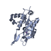
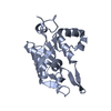
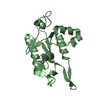
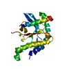
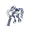
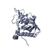
| Entry | Database: PDB / ID: 5h9v | ||||||
|---|---|---|---|---|---|---|---|
| Title | Crystal structure of Regnase PIN domain, form I | ||||||
 Components Components | Ribonuclease ZC3H12A | ||||||
 Keywords Keywords | HYDROLASE / RNase | ||||||
| Function / homology |  Function and homology information Function and homology informationimmune response-activating signaling pathway / positive regulation of protein deubiquitination / positive regulation of miRNA catabolic process / negative regulation of macrophage activation / miRNA catabolic process / negative regulation of muscle cell apoptotic process / RNA exonuclease activity / rough endoplasmic reticulum membrane / negative regulation of T-helper 17 cell differentiation / cellular response to sodium arsenite ...immune response-activating signaling pathway / positive regulation of protein deubiquitination / positive regulation of miRNA catabolic process / negative regulation of macrophage activation / miRNA catabolic process / negative regulation of muscle cell apoptotic process / RNA exonuclease activity / rough endoplasmic reticulum membrane / negative regulation of T-helper 17 cell differentiation / cellular response to sodium arsenite / host-mediated suppression of viral genome replication / positive regulation of mRNA catabolic process / cellular response to ionomycin / negative regulation of cardiac muscle contraction / 3'-UTR-mediated mRNA destabilization / nuclease activity / positive regulation of lipid storage / cellular response to chemokine / negative regulation of nitric oxide biosynthetic process / mRNA 3'-UTR AU-rich region binding / positive regulation of p38MAPK cascade / negative regulation of non-canonical NF-kappaB signal transduction / nuclear-transcribed mRNA catabolic process, nonsense-mediated decay / miRNA binding / negative regulation of interleukin-1 beta production / mRNA catabolic process / negative regulation of type II interferon production / protein complex oligomerization / negative regulation of interleukin-6 production / negative regulation of protein phosphorylation / negative regulation of tumor necrosis factor production / RNA nuclease activity / cellular response to interleukin-1 / positive regulation of execution phase of apoptosis / protein deubiquitination / positive regulation of fat cell differentiation / cellular response to glucose starvation / positive regulation of defense response to virus by host / rough endoplasmic reticulum / RNA endonuclease activity / negative regulation of cytokine production involved in inflammatory response / positive regulation of endothelial cell migration / positive regulation of autophagy / negative regulation of canonical NF-kappaB signal transduction / mRNA 3'-UTR binding / P-body / positive regulation of protein import into nucleus / cellular response to virus / RNA stem-loop binding / cellular response to tumor necrosis factor / positive regulation of reactive oxygen species metabolic process / positive regulation of angiogenesis / cytoplasmic ribonucleoprotein granule / T cell receptor signaling pathway / nervous system development / cellular response to lipopolysaccharide / regulation of gene expression / ribosome binding / cellular response to oxidative stress / angiogenesis / Hydrolases; Acting on ester bonds / cytoskeleton / cell differentiation / cysteine-type deubiquitinase activity / inflammatory response / negative regulation of gene expression / mRNA binding / chromatin binding / apoptotic process / positive regulation of gene expression / DNA damage response / negative regulation of transcription by RNA polymerase II / positive regulation of transcription by RNA polymerase II / protein-containing complex / DNA binding / RNA binding / zinc ion binding / nucleoplasm / nucleus / cytoplasm Similarity search - Function | ||||||
| Biological species |  | ||||||
| Method |  X-RAY DIFFRACTION / SYNCHROTRON / MAD / Resolution: 2.75 Å X-RAY DIFFRACTION / SYNCHROTRON / MAD / Resolution: 2.75 Å | ||||||
 Authors Authors | Yokogawa, M. / Tsushima, T. / Adachi, W. / Noda, N.N. / Inagaki, F. | ||||||
| Funding support |  Japan, 1items Japan, 1items
| ||||||
 Citation Citation | Journal: Sci Rep / Year: 2016 Title: Structural basis for the regulation of enzymatic activity of Regnase-1 by domain-domain interactions Authors: Yokogawa, M. / Tsushima, T. / Noda, N.N. / Kumeta, H. / Enokizono, Y. / Yamashita, K. / Standley, D.M. / Takeuchi, O. / Akira, S. / Inagaki, F. | ||||||
| History |
|
- Structure visualization
| Structure viewer | Molecule: MolmilJmol/JSmol |
|---|
- Downloads & links
-Download
| PDBx/mmCIF format | 5h9v.cif.gz | 284.6 KB | Display | PDBx/mmCIF format |
|---|---|---|---|---|
| PDB format | pdb5h9v.ent.gz | 233.6 KB | Display | PDB format |
| PDBx/mmJSON format | 5h9v.json.gz | Tree view | PDBx/mmJSON format | |
| Others |  Other downloads Other downloads |
-Validation report
| Arichive directory | https://data.pdbj.org/pub/pdb/validation_reports/h9/5h9vftp://data.pdbj.org/pub/pdb/validation_reports/h9/5h9v | HTTPS FTP |
|---|
-Related structure data


-Links
 PDBj
PDBj- Assembly
| Deposited unit | 
| |||||||||||||||||||||||||||||||||||||||||||||||||||||||||||||||||
|---|---|---|---|---|---|---|---|---|---|---|---|---|---|---|---|---|---|---|---|---|---|---|---|---|---|---|---|---|---|---|---|---|---|---|---|---|---|---|---|---|---|---|---|---|---|---|---|---|---|---|---|---|---|---|---|---|---|---|---|---|---|---|---|---|---|---|
| 1 | 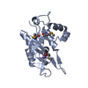
| |||||||||||||||||||||||||||||||||||||||||||||||||||||||||||||||||
| 2 | 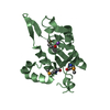
| |||||||||||||||||||||||||||||||||||||||||||||||||||||||||||||||||
| 3 | 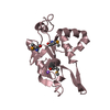
| |||||||||||||||||||||||||||||||||||||||||||||||||||||||||||||||||
| 4 | 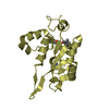
| |||||||||||||||||||||||||||||||||||||||||||||||||||||||||||||||||
| Unit cell |
| |||||||||||||||||||||||||||||||||||||||||||||||||||||||||||||||||
| Noncrystallographic symmetry (NCS) | NCS domain:
NCS domain segments:
NCS oper:
|
-Components
| #1: Protein | Mass: 24717.869 Da / Num. of mol.: 4 / Fragment: PIN domain, UNP residues 134-339 Source method: isolated from a genetically manipulated source Source: (gene. exp.)  References: UniProt: Q5D1E7, Hydrolases; Acting on ester bonds #2: Chemical | ChemComp-NA /   Mass: 22.990 Da / Num. of mol.: 4 / Source method: obtained synthetically / Formula: Na Mass: 22.990 Da / Num. of mol.: 4 / Source method: obtained synthetically / Formula: NaHas protein modification | Y | |
|---|
-Experimental details
-Experiment
| Experiment | Method: X-RAY DIFFRACTION |
|---|
- Sample preparation
| Crystal | Density Matthews: 3.52 Å3/Da / Density % sol: 65.03 % |
|---|---|
| Crystal grow | Temperature: 293 K / Method: vapor diffusion, sitting drop / pH: 5.5 / Details: 1 M (NH4)2HPO4, 200 mM NaCl, 100 mM sodium citrate |
-Data collection
| Diffraction | Mean temperature: 95 K | ||||||||||||
|---|---|---|---|---|---|---|---|---|---|---|---|---|---|
| Diffraction source | Source: SYNCHROTRON / Site: Photon Factory / Beamline: AR-NE3A / Wavelength: 0.97899, 0.97928, 0.96405 | ||||||||||||
| Detector | Type: ADSC QUANTUM 270 / Detector: CCD / Date: Apr 8, 2009 | ||||||||||||
| Radiation | Protocol: SINGLE WAVELENGTH / Monochromatic (M) / Laue (L): M / Scattering type: x-ray | ||||||||||||
| Radiation wavelength |
| ||||||||||||
| Reflection | Resolution: 2.75→47.49 Å / Num. obs: 33467 / % possible obs: 90.5 % / Redundancy: 7.9 % / Net I/σ(I): 22.2 |
- Processing
| Software |
| ||||||||||||||||||||||||||||||||||||||||||||||||||||||||||||||||||||||||||||||||||||||||||||||||||||||||||||||||||||||||||||||||||||||||||||||||||||||||||||||||||||||||||||||||||||||
|---|---|---|---|---|---|---|---|---|---|---|---|---|---|---|---|---|---|---|---|---|---|---|---|---|---|---|---|---|---|---|---|---|---|---|---|---|---|---|---|---|---|---|---|---|---|---|---|---|---|---|---|---|---|---|---|---|---|---|---|---|---|---|---|---|---|---|---|---|---|---|---|---|---|---|---|---|---|---|---|---|---|---|---|---|---|---|---|---|---|---|---|---|---|---|---|---|---|---|---|---|---|---|---|---|---|---|---|---|---|---|---|---|---|---|---|---|---|---|---|---|---|---|---|---|---|---|---|---|---|---|---|---|---|---|---|---|---|---|---|---|---|---|---|---|---|---|---|---|---|---|---|---|---|---|---|---|---|---|---|---|---|---|---|---|---|---|---|---|---|---|---|---|---|---|---|---|---|---|---|---|---|---|---|
| Refinement | Method to determine structure: MAD / Resolution: 2.75→47.49 Å / Cor.coef. Fo:Fc: 0.954 / Cor.coef. Fo:Fc free: 0.932 / SU B: 25.849 / SU ML: 0.22 / Cross valid method: THROUGHOUT / ESU R: 0.386 / ESU R Free: 0.267 / Stereochemistry target values: MAXIMUM LIKELIHOOD / Details: HYDROGENS HAVE BEEN ADDED IN THE RIDING POSITIONS
| ||||||||||||||||||||||||||||||||||||||||||||||||||||||||||||||||||||||||||||||||||||||||||||||||||||||||||||||||||||||||||||||||||||||||||||||||||||||||||||||||||||||||||||||||||||||
| Solvent computation | Ion probe radii: 0.8 Å / Shrinkage radii: 0.8 Å / VDW probe radii: 1.2 Å / Solvent model: MASK | ||||||||||||||||||||||||||||||||||||||||||||||||||||||||||||||||||||||||||||||||||||||||||||||||||||||||||||||||||||||||||||||||||||||||||||||||||||||||||||||||||||||||||||||||||||||
| Displacement parameters | Biso mean: 92.716 Å2
| ||||||||||||||||||||||||||||||||||||||||||||||||||||||||||||||||||||||||||||||||||||||||||||||||||||||||||||||||||||||||||||||||||||||||||||||||||||||||||||||||||||||||||||||||||||||
| Refinement step | Cycle: 1 / Resolution: 2.75→47.49 Å
| ||||||||||||||||||||||||||||||||||||||||||||||||||||||||||||||||||||||||||||||||||||||||||||||||||||||||||||||||||||||||||||||||||||||||||||||||||||||||||||||||||||||||||||||||||||||
| Refine LS restraints |
|