immune response-activating signaling pathway / positive regulation of protein deubiquitination / positive regulation of miRNA catabolic process / negative regulation of macrophage activation / miRNA catabolic process / negative regulation of muscle cell apoptotic process / RNA exonuclease activity / Regulation of CDH19 Expression and Function / rough endoplasmic reticulum membrane / negative regulation of T-helper 17 cell differentiation ...immune response-activating signaling pathway / positive regulation of protein deubiquitination / positive regulation of miRNA catabolic process / negative regulation of macrophage activation / miRNA catabolic process / negative regulation of muscle cell apoptotic process / RNA exonuclease activity / Regulation of CDH19 Expression and Function / rough endoplasmic reticulum membrane / negative regulation of T-helper 17 cell differentiation / cellular response to sodium arsenite / host-mediated suppression of viral genome replication / positive regulation of mRNA catabolic process / negative regulation of cardiac muscle contraction / cellular response to ionomycin / 3'-UTR-mediated mRNA destabilization / positive regulation of lipid storage / negative regulation of nitric oxide biosynthetic process / cellular response to chemokine / mRNA 3'-UTR AU-rich region binding / positive regulation of p38MAPK cascade / negative regulation of non-canonical NF-kappaB signal transduction / nuclear-transcribed mRNA catabolic process, nonsense-mediated decay / miRNA binding / negative regulation of interleukin-1 beta production / negative regulation of protein phosphorylation / negative regulation of type II interferon production / protein complex oligomerization / negative regulation of interleukin-6 production / negative regulation of tumor necrosis factor production / RNA nuclease activity / cellular response to interleukin-1 / positive regulation of execution phase of apoptosis / protein deubiquitination / positive regulation of fat cell differentiation / cellular response to glucose starvation / positive regulation of defense response to virus by host / RNA endonuclease activity / negative regulation of cytokine production involved in inflammatory response / positive regulation of endothelial cell migration / positive regulation of autophagy / negative regulation of canonical NF-kappaB signal transduction / mRNA 3'-UTR binding / P-body / cellular response to virus / positive regulation of protein import into nucleus / RNA stem-loop binding / cellular response to tumor necrosis factor / positive regulation of reactive oxygen species metabolic process / positive regulation of angiogenesis / cytoplasmic ribonucleoprotein granule / nervous system development / T cell receptor signaling pathway / cellular response to lipopolysaccharide / ribosome binding / regulation of gene expression / cellular response to oxidative stress / angiogenesis / defense response to virus / Hydrolases; Acting on ester bonds / cytoskeleton / cell differentiation / inflammatory response / mRNA binding / apoptotic process / chromatin binding / DNA damage response / positive regulation of gene expression / DNA-templated transcription / negative regulation of transcription by RNA polymerase II / positive regulation of transcription by RNA polymerase II / protein-containing complex / DNA binding / RNA binding / zinc ion binding / nucleoplasm / nucleus / cytoplasm Similarity search - Function
In the structure databanks used in Yorodumi, some data are registered as the other names, "COVID-19 virus" and "2019-nCoV". Here are the details of the virus and the list of structure data.
Jan 31, 2019. EMDB accession codes are about to change! (news from PDBe EMDB page)
EMDB accession codes are about to change! (news from PDBe EMDB page)
The allocation of 4 digits for EMDB accession codes will soon come to an end. Whilst these codes will remain in use, new EMDB accession codes will include an additional digit and will expand incrementally as the available range of codes is exhausted. The current 4-digit format prefixed with “EMD-” (i.e. EMD-XXXX) will advance to a 5-digit format (i.e. EMD-XXXXX), and so on. It is currently estimated that the 4-digit codes will be depleted around Spring 2019, at which point the 5-digit format will come into force.
The EM Navigator/Yorodumi systems omit the EMD- prefix.
Related info.:Q: What is EMD? / ID/Accession-code notation in Yorodumi/EM Navigator
Yorodumi is a browser for structure data from EMDB, PDB, SASBDB, etc.
This page is also the successor to EM Navigator detail page, and also detail information page/front-end page for Omokage search.
The word "yorodu" (or yorozu) is an old Japanese word meaning "ten thousand". "mi" (miru) is to see.
Related info.:EMDB / PDB / SASBDB / Comparison of 3 databanks / Yorodumi Search / Aug 31, 2016. New EM Navigator & Yorodumi / Yorodumi Papers / Jmol/JSmol / Function and homology information / Changes in new EM Navigator and Yorodumi
 Movie
Movie Controller

 Yorodumi
 Open data
 Basic information
 Components
 Keywords
 Function and homology information
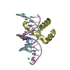
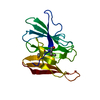
 Homo sapiens (human)
 X-RAY DIFFRACTION /
  Authors
 Citation
 Structure visualization
 Downloads & links
 Other downloads

 PDBj
PDBj Assembly


 Mass: 24.305 Da / Num. of mol.: 2 / Source method: obtained synthetically / Formula: Mg
 Mass: 18.015 Da / Num. of mol.: 157 / Source method: isolated from a natural source / Formula: H2O
 Sample preparation
 / Beamline: BL-17A / Wavelength: 1 Å
 Processing