 Movie
Movie Controller


+ Open data

- Basic information
| Entry | Database: PDB / ID: 7ndk | ||||||
|---|---|---|---|---|---|---|---|
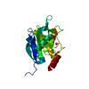
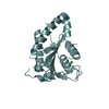
| Title | Crystal structure of ZC3H12C PIN catalytic mutant | ||||||
 Components Components | Probable ribonuclease ZC3H12C | ||||||
 Keywords Keywords | RNA BINDING PROTEIN / Endo-ribonuclease / Regnase | ||||||
| Function / homology |  Function and homology information Function and homology informationRNA endonuclease activity / cytoplasmic ribonucleoprotein granule / Hydrolases; Acting on ester bonds / hydrolase activity / mRNA binding / zinc ion binding / nucleus Similarity search - Function | ||||||
| Biological species |  | ||||||
| Method |  X-RAY DIFFRACTION / SYNCHROTRON / MOLECULAR REPLACEMENT / Resolution: 2.34 Å X-RAY DIFFRACTION / SYNCHROTRON / MOLECULAR REPLACEMENT / Resolution: 2.34 Å | ||||||
 Authors Authors | Garg, A. / Heinemann, U. | ||||||
 Citation Citation | Journal: Nucleic Acids Res. / Year: 2021 Title: PIN and CCCH Zn-finger domains coordinate RNA targeting in ZC3H12 family endoribonucleases. Authors: Garg, A. / Roske, Y. / Yamada, S. / Uehata, T. / Takeuchi, O. / Heinemann, U. | ||||||
| History |
|
- Structure visualization
| Structure viewer | Molecule: MolmilJmol/JSmol |
|---|
- Downloads & links
-Download
| PDBx/mmCIF format | 7ndk.cif.gz | 139.9 KB | Display | PDBx/mmCIF format |
|---|---|---|---|---|
| PDB format | pdb7ndk.ent.gz | 109.7 KB | Display | PDB format |
| PDBx/mmJSON format | 7ndk.json.gz | Tree view | PDBx/mmJSON format | |
| Others |  Other downloads Other downloads |
-Validation report
| Arichive directory | https://data.pdbj.org/pub/pdb/validation_reports/nd/7ndkftp://data.pdbj.org/pub/pdb/validation_reports/nd/7ndk | HTTPS FTP |
|---|
-Related structure data
| Related structure data | 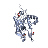 7ndhC 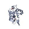 7ndiSC 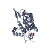 7ndjC S: Starting model for refinement C: citing same article ( |
|---|---|
| Similar structure data |


-Links
 PDBj
PDBj
- Assembly
| Deposited unit | 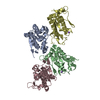
| ||||||||
|---|---|---|---|---|---|---|---|---|---|
| 1 | 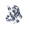
| ||||||||
| 2 | 
| ||||||||
| 3 | 
| ||||||||
| 4 | 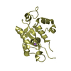
| ||||||||
| Unit cell |
|
-Components
| #1: Protein | Mass: 19151.951 Da / Num. of mol.: 4 Source method: isolated from a genetically manipulated source Source: (gene. exp.)  References: UniProt: Q5DTV4, Hydrolases; Acting on ester bonds #2: Chemical |   Mass: 22.990 Da / Num. of mol.: 2 / Source method: obtained synthetically / Formula: Na Mass: 22.990 Da / Num. of mol.: 2 / Source method: obtained synthetically / Formula: Na#3: Water | ChemComp-HOH / |  Mass: 18.015 Da / Num. of mol.: 94 / Source method: isolated from a natural source / Formula: H2O Mass: 18.015 Da / Num. of mol.: 94 / Source method: isolated from a natural source / Formula: H2OHas ligand of interest | N | |
|---|
-Experimental details
-Experiment
| Experiment | Method: X-RAY DIFFRACTION / Number of used crystals: 1 |
|---|
- Sample preparation
| Crystal grow | Temperature: 293 K / Method: vapor diffusion, sitting drop Details: 0.2 M KSCN, 20% PEG 3350 and 0.1 M Bis-Tris propane pH 6.5 |
|---|
-Data collection
| Diffraction | Mean temperature: 100 K / Serial crystal experiment: N | |||||||||||||||
|---|---|---|---|---|---|---|---|---|---|---|---|---|---|---|---|---|
| Diffraction source | Source: SYNCHROTRON / Site: BESSY  / Beamline: 14.1 / Wavelength: 0.91841 Å / Beamline: 14.1 / Wavelength: 0.91841 Å | |||||||||||||||
| Detector | Type: DECTRIS PILATUS3 S 6M / Detector: PIXEL / Date: Nov 2, 2016 | |||||||||||||||
| Radiation | Protocol: SINGLE WAVELENGTH / Monochromatic (M) / Laue (L): M / Scattering type: x-ray | |||||||||||||||
| Radiation wavelength | Wavelength: 0.91841 Å / Relative weight: 1 | |||||||||||||||
| Reflection twin |
| |||||||||||||||
| Reflection | Resolution: 2.34→38.75 Å / Num. obs: 21746 / % possible obs: 97 % / Redundancy: 2.25 % / CC1/2: 0.998 / Net I/σ(I): 10.19 | |||||||||||||||
| Reflection shell | Resolution: 2.34→2.39 Å / Num. unique obs: 1417 / CC1/2: 0.629 |
- Processing
| Software |
| ||||||||||||||||||||||||||||||||||||||||||||||||||||||||||||
|---|---|---|---|---|---|---|---|---|---|---|---|---|---|---|---|---|---|---|---|---|---|---|---|---|---|---|---|---|---|---|---|---|---|---|---|---|---|---|---|---|---|---|---|---|---|---|---|---|---|---|---|---|---|---|---|---|---|---|---|---|---|
| Refinement | Method to determine structure: MOLECULAR REPLACEMENT Starting model: 7NDI Resolution: 2.34→38.75 Å / Cor.coef. Fo:Fc: 0.942 / Cor.coef. Fo:Fc free: 0.921 / SU B: 7.299 / SU ML: 0.189 / Cross valid method: THROUGHOUT / σ(F): 0 / ESU R Free: 0.07 / Stereochemistry target values: MAXIMUM LIKELIHOOD
| ||||||||||||||||||||||||||||||||||||||||||||||||||||||||||||
| Solvent computation | Ion probe radii: 0.8 Å / Shrinkage radii: 0.8 Å / VDW probe radii: 1.2 Å / Solvent model: MASK | ||||||||||||||||||||||||||||||||||||||||||||||||||||||||||||
| Displacement parameters | Biso max: 141.13 Å2 / Biso mean: 61.591 Å2 / Biso min: 27.73 Å2
| ||||||||||||||||||||||||||||||||||||||||||||||||||||||||||||
| Refinement step | Cycle: final / Resolution: 2.34→38.75 Å
| ||||||||||||||||||||||||||||||||||||||||||||||||||||||||||||
| Refine LS restraints |
| ||||||||||||||||||||||||||||||||||||||||||||||||||||||||||||
| LS refinement shell | Resolution: 2.34→2.398 Å / Rfactor Rfree error: 0
|
