 Movie
Movie Controller


+ Open data

- Basic information
| Entry | Database: PDB / ID: 7m1c | ||||||
|---|---|---|---|---|---|---|---|
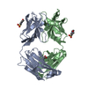
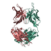
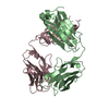
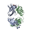
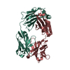
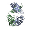
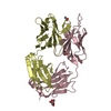
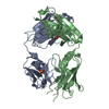
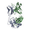
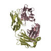
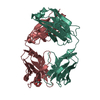
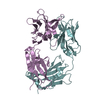
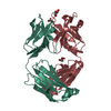
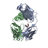
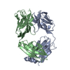
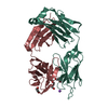
| Title | Crystal structure of the HCMV pentamer-specific antibody 1-32 | ||||||
 Components Components |
| ||||||
 Keywords Keywords | IMMUNE SYSTEM / HCMV pentamer / Fab / cytomegalovirus / antibody | ||||||
| Function / homology | Immunoglobulins / Immunoglobulin-like / Sandwich / Mainly Beta Function and homology information Function and homology information | ||||||
| Biological species |  Homo sapiens (human) Homo sapiens (human) | ||||||
| Method |  X-RAY DIFFRACTION / SYNCHROTRON / MOLECULAR REPLACEMENT / Resolution: 2.1 Å X-RAY DIFFRACTION / SYNCHROTRON / MOLECULAR REPLACEMENT / Resolution: 2.1 Å | ||||||
 Authors Authors | Wrapp, D. / McLellan, J.S. | ||||||
 Citation Citation | Journal: Sci Adv / Year: 2022 Title: Structural basis for HCMV Pentamer recognition by neuropilin 2 and neutralizing antibodies. Authors: Daniel Wrapp / Xiaohua Ye / Zhiqiang Ku / Hang Su / Harrison G Jones / Nianshuang Wang / Akaash K Mishra / Daniel C Freed / Fengsheng Li / Aimin Tang / Leike Li / Dabbu Kumar Jaijyan / Hua ...Authors: Daniel Wrapp / Xiaohua Ye / Zhiqiang Ku / Hang Su / Harrison G Jones / Nianshuang Wang / Akaash K Mishra / Daniel C Freed / Fengsheng Li / Aimin Tang / Leike Li / Dabbu Kumar Jaijyan / Hua Zhu / Dai Wang / Tong-Ming Fu / Ningyan Zhang / Zhiqiang An / Jason S McLellan /  Abstract: Human cytomegalovirus (HCMV) encodes multiple surface glycoprotein complexes to infect a variety of cell types. The HCMV Pentamer, composed of gH, gL, UL128, UL130, and UL131A, enhances entry into ...Human cytomegalovirus (HCMV) encodes multiple surface glycoprotein complexes to infect a variety of cell types. The HCMV Pentamer, composed of gH, gL, UL128, UL130, and UL131A, enhances entry into epithelial, endothelial, and myeloid cells by interacting with the cell surface receptor neuropilin 2 (NRP2). Despite the critical nature of this interaction, the molecular determinants that govern NRP2 recognition remain unclear. Here, we describe the cryo-EM structure of NRP2 bound to Pentamer. The high-affinity interaction between these proteins is calcium dependent and differs from the canonical carboxyl-terminal arginine (CendR) binding that NRP2 typically uses. We also determine the structures of four neutralizing human antibodies bound to the HCMV Pentamer to define susceptible epitopes. Two of these antibodies compete with NRP2 binding, but the two most potent antibodies recognize a previously unidentified epitope that does not overlap the NRP2-binding site. Collectively, these findings provide a structural basis for HCMV tropism and antibody-mediated neutralization. | ||||||
| History |
|
- Structure visualization
| Structure viewer | Molecule: MolmilJmol/JSmol |
|---|
- Downloads & links
-Download
| PDBx/mmCIF format | 7m1c.cif.gz | 179.5 KB | Display | PDBx/mmCIF format |
|---|---|---|---|---|
| PDB format | pdb7m1c.ent.gz | 142.4 KB | Display | PDB format |
| PDBx/mmJSON format | 7m1c.json.gz | Tree view | PDBx/mmJSON format | |
| Others |  Other downloads Other downloads |
-Validation report
| Arichive directory | https://data.pdbj.org/pub/pdb/validation_reports/m1/7m1cftp://data.pdbj.org/pub/pdb/validation_reports/m1/7m1c | HTTPS FTP |
|---|
-Related structure data
| Related structure data | 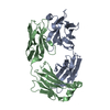 7kbaC 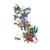 7kbbC 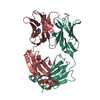 7lyvC 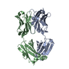 7lywC  7m22C  7m30C  3u2sS S: Starting model for refinement C: citing same article ( |
|---|---|
| Similar structure data |


-Links
 PDBj
PDBj

- Assembly
| Deposited unit | 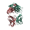
| ||||||||
|---|---|---|---|---|---|---|---|---|---|
| 1 |
| ||||||||
| Unit cell |
|
-Components
| #1: Antibody | Mass: 26082.371 Da / Num. of mol.: 1 Source method: isolated from a genetically manipulated source Source: (gene. exp.) Homo sapiens (human) / Production host: Homo sapiens (human) |
|---|---|
| #2: Antibody | Mass: 23319.729 Da / Num. of mol.: 1 Source method: isolated from a genetically manipulated source Source: (gene. exp.) Homo sapiens (human) / Production host: Homo sapiens (human) |
| #3: Water | ChemComp-HOH /  Mass: 18.015 Da / Num. of mol.: 70 / Source method: isolated from a natural source / Formula: H2O Mass: 18.015 Da / Num. of mol.: 70 / Source method: isolated from a natural source / Formula: H2O |
| Has protein modification | Y |
-Experimental details
-Experiment
| Experiment | Method: X-RAY DIFFRACTION / Number of used crystals: 1 |
|---|
- Sample preparation
| Crystal | Density Matthews: 2.09 Å3/Da / Density % sol: 41.27 % |
|---|---|
| Crystal grow | Temperature: 293.15 K / Method: vapor diffusion, hanging drop Details: 2.0 M ammonium sulfate, 0.2 M sodium chloride, 5% isopropanol |
-Data collection
| Diffraction | Mean temperature: 80 K / Serial crystal experiment: N |
|---|---|
| Diffraction source | Source: SYNCHROTRON / Site: APS / Beamline: 19-ID / Wavelength: 0.979 Å |
| Detector | Type: ADSC QUANTUM 315r / Detector: CCD / Date: Aug 1, 2019 |
| Radiation | Protocol: SINGLE WAVELENGTH / Monochromatic (M) / Laue (L): M / Scattering type: x-ray |
| Radiation wavelength | Wavelength: 0.979 Å / Relative weight: 1 |
| Reflection | Resolution: 2.1→54.2 Å / Num. obs: 25404 / % possible obs: 99.9 % / Redundancy: 7.2 % / CC1/2: 0.987 / Net I/σ(I): 6 |
| Reflection shell | Resolution: 2.1→2.16 Å / Mean I/σ(I) obs: 1.5 / Num. unique obs: 13538 / CC1/2: 0.753 / % possible all: 99.8 |
- Processing
| Software |
| ||||||||||||||||||
|---|---|---|---|---|---|---|---|---|---|---|---|---|---|---|---|---|---|---|---|
| Refinement | Method to determine structure: MOLECULAR REPLACEMENT Starting model: 3U2S Resolution: 2.1→47.98 Å / Cross valid method: FREE R-VALUE
| ||||||||||||||||||
| Displacement parameters | Biso max: 119.85 Å2 / Biso mean: 62.57 Å2 / Biso min: 29.98 Å2 | ||||||||||||||||||
| Refinement step | Cycle: LAST / Resolution: 2.1→47.98 Å
|
