 Movie
Movie Controller

[English] 日本語
 Yorodumi
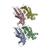
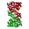
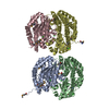
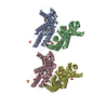
- PDB-7kbb: Cryo-EM structure of the HCMV pentamer bound by Fabs 2-18 and 8I21 -
+ Open data

- Basic information
| Entry | Database: PDB / ID: 7kbb | ||||||
|---|---|---|---|---|---|---|---|
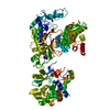
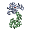
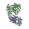
| Title | Cryo-EM structure of the HCMV pentamer bound by Fabs 2-18 and 8I21 | ||||||
 Components Components |
| ||||||
 Keywords Keywords | VIRAL PROTEIN/IMMUNE SYSTEM / HCMV pentamer / Fab / complex / VIRAL PROTEIN / VIRAL PROTEIN-IMMUNE SYSTEM complex | ||||||
| Function / homology | Herpesvirus UL130, cytomegalovirus / : / HCMV glycoprotein pUL130 / Cytomegalovirus UL128 protein / viral envelope / Envelope glycoprotein UL130 / UL128 / UL131A Function and homology information Function and homology information | ||||||
| Biological species |  Homo sapiens (human) Homo sapiens (human)  Human cytomegalovirus Human cytomegalovirus | ||||||
| Method | ELECTRON MICROSCOPY / single particle reconstruction / cryo EM / Resolution: 4.02 Å | ||||||
 Authors Authors | Wrapp, D. / McLellan, J.S. | ||||||
 Citation Citation | Journal: Sci Adv / Year: 2022 Title: Structural basis for HCMV Pentamer recognition by neuropilin 2 and neutralizing antibodies. Authors: Daniel Wrapp / Xiaohua Ye / Zhiqiang Ku / Hang Su / Harrison G Jones / Nianshuang Wang / Akaash K Mishra / Daniel C Freed / Fengsheng Li / Aimin Tang / Leike Li / Dabbu Kumar Jaijyan / Hua ...Authors: Daniel Wrapp / Xiaohua Ye / Zhiqiang Ku / Hang Su / Harrison G Jones / Nianshuang Wang / Akaash K Mishra / Daniel C Freed / Fengsheng Li / Aimin Tang / Leike Li / Dabbu Kumar Jaijyan / Hua Zhu / Dai Wang / Tong-Ming Fu / Ningyan Zhang / Zhiqiang An / Jason S McLellan /  Abstract: Human cytomegalovirus (HCMV) encodes multiple surface glycoprotein complexes to infect a variety of cell types. The HCMV Pentamer, composed of gH, gL, UL128, UL130, and UL131A, enhances entry into ...Human cytomegalovirus (HCMV) encodes multiple surface glycoprotein complexes to infect a variety of cell types. The HCMV Pentamer, composed of gH, gL, UL128, UL130, and UL131A, enhances entry into epithelial, endothelial, and myeloid cells by interacting with the cell surface receptor neuropilin 2 (NRP2). Despite the critical nature of this interaction, the molecular determinants that govern NRP2 recognition remain unclear. Here, we describe the cryo-EM structure of NRP2 bound to Pentamer. The high-affinity interaction between these proteins is calcium dependent and differs from the canonical carboxyl-terminal arginine (CendR) binding that NRP2 typically uses. We also determine the structures of four neutralizing human antibodies bound to the HCMV Pentamer to define susceptible epitopes. Two of these antibodies compete with NRP2 binding, but the two most potent antibodies recognize a previously unidentified epitope that does not overlap the NRP2-binding site. Collectively, these findings provide a structural basis for HCMV tropism and antibody-mediated neutralization. | ||||||
| History |
|
- Structure visualization
| Movie |
Movie viewer |
|---|---|
| Structure viewer | Molecule: MolmilJmol/JSmol |
- Downloads & links
-Download
| PDBx/mmCIF format | 7kbb.cif.gz | 181.8 KB | Display | PDBx/mmCIF format |
|---|---|---|---|---|
| PDB format | pdb7kbb.ent.gz | 131.4 KB | Display | PDB format |
| PDBx/mmJSON format | 7kbb.json.gz | Tree view | PDBx/mmJSON format | |
| Others |  Other downloads Other downloads |
-Validation report
| Arichive directory | https://data.pdbj.org/pub/pdb/validation_reports/kb/7kbbftp://data.pdbj.org/pub/pdb/validation_reports/kb/7kbb | HTTPS FTP |
|---|
-Related structure data
| Related structure data |  22788MC 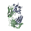 7kbaC 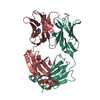 7lyvC 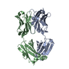 7lywC 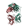 7m1cC 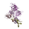 7m22C  7m30C M: map data used to model this data C: citing same article ( |
|---|---|
| Similar structure data |


-Links
 PDBj
PDBj

- Assembly
| Deposited unit | 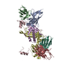
|
|---|---|
| 1 |
|
-Components
-Protein , 3 types, 3 molecules CDE
| #3: Protein | Mass: 16684.299 Da / Num. of mol.: 1 Source method: isolated from a genetically manipulated source Source: (gene. exp.) Human cytomegalovirus / Gene: UL128 / Production host: Homo sapiens (human) / References: UniProt: C8BLJ3 |
|---|---|
| #4: Protein | Mass: 21761.678 Da / Num. of mol.: 1 Source method: isolated from a genetically manipulated source Source: (gene. exp.) Human cytomegalovirus / Gene: UL130 / Production host: Homo sapiens (human) / References: UniProt: A0A0G2TB82 |
| #5: Protein | Mass: 13005.457 Da / Num. of mol.: 1 Source method: isolated from a genetically manipulated source Source: (gene. exp.) Human cytomegalovirus / Production host: Homo sapiens (human) / References: UniProt: Q38M21 |
-Antibody , 4 types, 4 molecules ABHL
| #1: Antibody | Mass: 25640.506 Da / Num. of mol.: 1 Source method: isolated from a genetically manipulated source Source: (gene. exp.) Homo sapiens (human) / Production host: Homo sapiens (human) |
|---|---|
| #2: Antibody | Mass: 23414.055 Da / Num. of mol.: 1 Source method: isolated from a genetically manipulated source Source: (gene. exp.) Homo sapiens (human) / Production host: Homo sapiens (human) |
| #6: Antibody | Mass: 25196.340 Da / Num. of mol.: 1 Source method: isolated from a genetically manipulated source Source: (gene. exp.) Homo sapiens (human) / Production host: Homo sapiens (human) |
| #7: Antibody | Mass: 23459.006 Da / Num. of mol.: 1 Source method: isolated from a genetically manipulated source Source: (gene. exp.) Homo sapiens (human) / Production host: Homo sapiens (human) |
-Details
| Has protein modification | Y |
|---|
-Experimental details
-Experiment
| Experiment | Method: ELECTRON MICROSCOPY |
|---|---|
| EM experiment | Aggregation state: PARTICLE / 3D reconstruction method: single particle reconstruction |
- Sample preparation
| Component |
| ||||||||||||||||||||||||||||||
|---|---|---|---|---|---|---|---|---|---|---|---|---|---|---|---|---|---|---|---|---|---|---|---|---|---|---|---|---|---|---|---|
| Molecular weight | Value: 0.28 MDa / Experimental value: YES | ||||||||||||||||||||||||||||||
| Source (natural) |
| ||||||||||||||||||||||||||||||
| Source (recombinant) |
| ||||||||||||||||||||||||||||||
| Buffer solution | pH: 8 | ||||||||||||||||||||||||||||||
| Buffer component |
| ||||||||||||||||||||||||||||||
| Specimen | Conc.: 0.25 mg/ml / Embedding applied: NO / Shadowing applied: NO / Staining applied: NO / Vitrification applied: YES | ||||||||||||||||||||||||||||||
| Specimen support | Grid material: COPPER / Grid mesh size: 400 divisions/in. / Grid type: C-flat-1.2/1.3 | ||||||||||||||||||||||||||||||
| Vitrification | Instrument: FEI VITROBOT MARK IV / Cryogen name: ETHANE / Humidity: 100 % / Chamber temperature: 277 K |
- Electron microscopy imaging
| Experimental equipment |  Model: Titan Krios / Image courtesy: FEI Company |
|---|---|
| Microscopy | Model: FEI TITAN KRIOS |
| Electron gun | Electron source:  FIELD EMISSION GUN / Accelerating voltage: 300 kV / Illumination mode: FLOOD BEAM FIELD EMISSION GUN / Accelerating voltage: 300 kV / Illumination mode: FLOOD BEAM |
| Electron lens | Mode: BRIGHT FIELD / C2 aperture diameter: 100 µm |
| Image recording | Electron dose: 36 e/Å2 / Film or detector model: GATAN K3 (6k x 4k) |
- Processing
| Software | Name: PHENIX / Classification: refinement | ||||||||||||||||||
|---|---|---|---|---|---|---|---|---|---|---|---|---|---|---|---|---|---|---|---|
| EM software |
| ||||||||||||||||||
| CTF correction | Type: PHASE FLIPPING AND AMPLITUDE CORRECTION | ||||||||||||||||||
| Symmetry | Point symmetry: C1 (asymmetric) | ||||||||||||||||||
| 3D reconstruction | Resolution: 4.02 Å / Resolution method: FSC 0.143 CUT-OFF / Num. of particles: 249802 / Symmetry type: POINT | ||||||||||||||||||
| Refinement | Cross valid method: THROUGHOUT | ||||||||||||||||||
| Displacement parameters | Biso max: 102.73 Å2 / Biso mean: 57.7257 Å2 / Biso min: 30 Å2 |
