 Movie
Movie Controller

[English] 日本語
 Yorodumi
- PDB-2ad5: Mechanisms of feedback regulation and drug resistance of CTP synt... -
+ Open data

- Basic information
| Entry | Database: PDB / ID: 2ad5 | ||||||
|---|---|---|---|---|---|---|---|
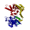
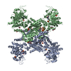
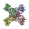
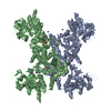
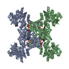
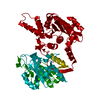
| Title | Mechanisms of feedback regulation and drug resistance of CTP synthetases: structure of the E. coli CTPS/CTP complex at 2.8-Angstrom resolution. | ||||||
 Components Components | CTP synthase | ||||||
 Keywords Keywords | LIGASE / Rossmann fold / P-loop ATPase / interfacial active site | ||||||
| Function / homology |  Function and homology information Function and homology informationCTP synthase (glutamine hydrolysing) / CTP synthase activity / 'de novo' CTP biosynthetic process / cytoophidium / pyrimidine nucleobase biosynthetic process / glutaminase activity / CTP biosynthetic process / protein homotetramerization / magnesium ion binding / protein-containing complex ...CTP synthase (glutamine hydrolysing) / CTP synthase activity / 'de novo' CTP biosynthetic process / cytoophidium / pyrimidine nucleobase biosynthetic process / glutaminase activity / CTP biosynthetic process / protein homotetramerization / magnesium ion binding / protein-containing complex / ATP binding / identical protein binding / cytosol Similarity search - Function | ||||||
| Biological species |  | ||||||
| Method |  X-RAY DIFFRACTION / SYNCHROTRON / FOURIER SYNTHESIS / Resolution: 2.8 Å X-RAY DIFFRACTION / SYNCHROTRON / FOURIER SYNTHESIS / Resolution: 2.8 Å | ||||||
 Authors Authors | Endrizzi, J.A. / Kim, H. / Anderson, P.M. / Baldwin, E.P. | ||||||
 Citation Citation | Journal: Biochemistry / Year: 2005 Title: Mechanisms of Product Feedback Regulation and Drug Resistance in Cytidine Triphosphate Synthetases from the Structure of a CTP-Inhibited Complex(,). Authors: Endrizzi, J.A. / Kim, H. / Anderson, P.M. / Baldwin, E.P. #1: Journal: Biochemistry / Year: 2004Title: Crystal structure of Escherichia coli cytidine triphosphate synthetase, a nucleotide-regulated glutamine amidotransferase/ATP-dependent amidoligase fusion protein and homologue of anticancer ...Title: Crystal structure of Escherichia coli cytidine triphosphate synthetase, a nucleotide-regulated glutamine amidotransferase/ATP-dependent amidoligase fusion protein and homologue of anticancer and antiparasitic drug targets. Authors: Endrizzi, J.A. / Kim, H. / Anderson, P.M. / Baldwin, E.P. | ||||||
| History |
|
- Structure visualization
| Structure viewer | Molecule: MolmilJmol/JSmol |
|---|
- Downloads & links
-Download
| PDBx/mmCIF format | 2ad5.cif.gz | 227.8 KB | Display | PDBx/mmCIF format |
|---|---|---|---|---|
| PDB format | pdb2ad5.ent.gz | 179.9 KB | Display | PDB format |
| PDBx/mmJSON format | 2ad5.json.gz | Tree view | PDBx/mmJSON format | |
| Others |  Other downloads Other downloads |
-Validation report
| Arichive directory | https://data.pdbj.org/pub/pdb/validation_reports/ad/2ad5ftp://data.pdbj.org/pub/pdb/validation_reports/ad/2ad5 | HTTPS FTP |
|---|
-Related structure data
| Related structure data |  1s1mS S: Starting model for refinement |
|---|---|
| Similar structure data |

-Links
 PDBj
PDBj

- Assembly
| Deposited unit | 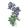
| ||||||||
|---|---|---|---|---|---|---|---|---|---|
| 1 | 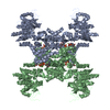
| ||||||||
| 2 | 
| ||||||||
| 3 | 
| ||||||||
| Unit cell |
| ||||||||
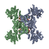
| Details | The tetrameric biological assembly is generated from the asymmetric unit dimer by the crystallographic two-fold axis: -x, -y, z |
-Components
| #1: Protein | Mass: 60446.980 Da / Num. of mol.: 2 / Source method: isolated from a natural source / Details: soluble cytoplasmic / Source: (natural) References: UniProt: P0A7E5, CTP synthase (glutamine hydrolysing) #2: Chemical |   Mass: 24.305 Da / Num. of mol.: 2 / Source method: obtained synthetically / Formula: Mg Mass: 24.305 Da / Num. of mol.: 2 / Source method: obtained synthetically / Formula: Mg#3: Chemical |   Mass: 427.201 Da / Num. of mol.: 2 / Source method: obtained synthetically / Formula: C10H15N5O10P2 / Comment: ADP, energy-carrying molecule*YM Mass: 427.201 Da / Num. of mol.: 2 / Source method: obtained synthetically / Formula: C10H15N5O10P2 / Comment: ADP, energy-carrying molecule*YM#4: Chemical |   Mass: 483.156 Da / Num. of mol.: 2 / Source method: obtained synthetically / Formula: C9H16N3O14P3 Mass: 483.156 Da / Num. of mol.: 2 / Source method: obtained synthetically / Formula: C9H16N3O14P3#5: Water | ChemComp-HOH / |  Mass: 18.015 Da / Num. of mol.: 252 / Source method: isolated from a natural source / Formula: H2O Mass: 18.015 Da / Num. of mol.: 252 / Source method: isolated from a natural source / Formula: H2OHas protein modification | Y | |
|---|
-Experimental details
-Experiment
| Experiment | Method: X-RAY DIFFRACTION / Number of used crystals: 1 |
|---|
- Sample preparation
| Crystal | Density Matthews: 4.6 Å3/Da / Density % sol: 73 % |
|---|---|
| Crystal grow | Temperature: 277 K / Method: vapor diffusion, hanging drop / pH: 8.5 Details: Tris-Cl, ammonium sulfate, pH 8.5, VAPOR DIFFUSION, HANGING DROP, temperature 277K |
-Data collection
| Diffraction | Mean temperature: 100 K |
|---|---|
| Diffraction source | Source: SYNCHROTRON / Site: SSRL  / Beamline: BL7-1 / Wavelength: 0.975 Å / Beamline: BL7-1 / Wavelength: 0.975 Å |
| Detector | Type: MARRESEARCH / Detector: IMAGE PLATE / Date: Jun 13, 2004 |
| Radiation | Protocol: SINGLE WAVELENGTH / Monochromatic (M) / Laue (L): M / Scattering type: x-ray |
| Radiation wavelength | Wavelength: 0.975 Å / Relative weight: 1 |
| Reflection | Resolution: 2.8→45.4 Å / Num. all: 55879 / Num. obs: 55879 / % possible obs: 98.9 % / Observed criterion σ(F): -3 / Observed criterion σ(I): -3 / Redundancy: 3.3 % / Biso Wilson estimate: 45 Å2 / Rmerge(I) obs: 0.084 / Rsym value: 0.084 / Net I/σ(I): 8.5 |
| Reflection shell | Resolution: 2.8→2.95 Å / Redundancy: 3 % / Rmerge(I) obs: 0.31 / Mean I/σ(I) obs: 2.6 / Rsym value: 0.31 / % possible all: 95.1 |
- Processing
| Software |
| ||||||||||||||||||||
|---|---|---|---|---|---|---|---|---|---|---|---|---|---|---|---|---|---|---|---|---|---|
| Refinement | Method to determine structure: FOURIER SYNTHESIS Starting model: PDB entyry 1S1M Resolution: 2.8→20 Å / Isotropic thermal model: Isotropic / Cross valid method: THROUGHOUT / σ(F): -3 / Stereochemistry target values: Engh & Huber
| ||||||||||||||||||||
| Displacement parameters | Biso mean: 60 Å2
| ||||||||||||||||||||
| Refinement step | Cycle: LAST / Resolution: 2.8→20 Å
| ||||||||||||||||||||
| Refine LS restraints |
| ||||||||||||||||||||
| LS refinement shell | Resolution: 2.8→2.95 Å
|
