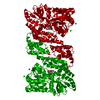
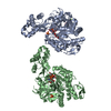
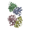
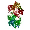
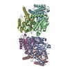
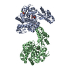
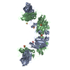
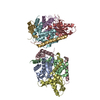
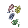
D-family DNA polymerase - DP2 subunit (catalytic subunit)
Components
DNA polymerase II large subunit
Keywords
TRANSFERASE / DNA polymerase D-family
Function / homology
Function and homology information
exodeoxyribonuclease I / intein-mediated protein splicing / single-stranded DNA 3'-5' DNA exonuclease activity / DNA catabolic process / DNA-templated DNA replication / DNA-directed DNA polymerase / DNA-directed DNA polymerase activity / DNA binding Similarity search - Function
DNA polymerase II large subunit DP2 / DNA polymerase II large subunit DP2, N-terminal / : / : / DNA polymerase II large subunit DP2, N-terminal / DNA polymerase II large subunit DP2, central domain / DNA polymerase II large subunit DP2, catalytic domain / Intein splicing domain / Intein C-terminal splicing region / Intein C-terminal splicing motif profile. ...DNA polymerase II large subunit DP2 / DNA polymerase II large subunit DP2, N-terminal / : / : / DNA polymerase II large subunit DP2, N-terminal / DNA polymerase II large subunit DP2, central domain / DNA polymerase II large subunit DP2, catalytic domain / Intein splicing domain / Intein C-terminal splicing region / Intein C-terminal splicing motif profile. / Intein N-terminal splicing region / Intein N-terminal splicing motif profile. / Hint domain N-terminal / Hint (Hedgehog/Intein) domain N-terminal region / Hint domain superfamily Similarity search - Domain/homology
Protocol: SINGLE WAVELENGTH / Monochromatic (M) / Laue (L): M / Scattering type: x-ray
Radiation wavelength
Wavelength: 0.984 Å / Relative weight: 1
Reflection
Resolution: 2.19→43.8 Å / Num. obs: 63302 / % possible obs: 99.6 % / Redundancy: 5.5 % / Biso Wilson estimate: 49.98 Å2 / Net I/σ(I): 8.5
Reflection shell
Rsym value: 1.151
-
Processing
Software
Name
Version
Classification
BUSTER
2.10.2
refinement
XDS
datareduction
Aimless
datascaling
AutoSol
phasing
Refinement
Method to determine structure: SAD / Resolution: 2.19→43.8 Å / Cor.coef. Fo:Fc: 0.9432 / Cor.coef. Fo:Fc free: 0.9278 / SU R Cruickshank DPI: 0.214 / Cross valid method: THROUGHOUT / σ(F): 0 / SU R Blow DPI: 0.212 / SU Rfree Blow DPI: 0.178 / SU Rfree Cruickshank DPI: 0.18
Rfactor
Num. reflection
% reflection
Selection details
Rfree
0.2346
3165
5 %
RANDOM
Rwork
0.199
-
-
-
obs
0.2008
63302
99.34 %
-
Displacement parameters
Biso mean: 53.38 Å2
Baniso -1
Baniso -2
Baniso -3
1-
-7.5119 Å2
0 Å2
0 Å2
2-
-
3.9661 Å2
0 Å2
3-
-
-
3.5458 Å2
Refine analyze
Luzzati coordinate error obs: 0.309 Å
Refinement step
Cycle: LAST / Resolution: 2.19→43.8 Å
Protein
Nucleic acid
Ligand
Solvent
Total
Num. atoms
7525
0
2
242
7769
Refine LS restraints
Refine-ID
Type
Dev ideal
Number
Restraint function
Weight
X-RAY DIFFRACTION
t_bond_d
0.01
7693
HARMONIC
2
X-RAY DIFFRACTION
t_angle_deg
1.09
10407
HARMONIC
2
X-RAY DIFFRACTION
t_dihedral_angle_d
2709
SINUSOIDAL
2
X-RAY DIFFRACTION
t_incorr_chiral_ct
X-RAY DIFFRACTION
t_pseud_angle
X-RAY DIFFRACTION
t_trig_c_planes
189
HARMONIC
2
X-RAY DIFFRACTION
t_gen_planes
1095
HARMONIC
5
X-RAY DIFFRACTION
t_it
7693
HARMONIC
20
X-RAY DIFFRACTION
t_nbd
0
SEMIHARMONIC
5
X-RAY DIFFRACTION
t_omega_torsion
3.08
X-RAY DIFFRACTION
t_other_torsion
18.25
X-RAY DIFFRACTION
t_improper_torsion
X-RAY DIFFRACTION
t_chiral_improper_torsion
965
SEMIHARMONIC
5
X-RAY DIFFRACTION
t_sum_occupancies
X-RAY DIFFRACTION
t_utility_distance
8
HARMONIC
1
X-RAY DIFFRACTION
t_utility_angle
12
HARMONIC
1
X-RAY DIFFRACTION
t_utility_torsion
X-RAY DIFFRACTION
t_ideal_dist_contact
8589
SEMIHARMONIC
4
LS refinement shell
Resolution: 2.19→2.25 Å / Total num. of bins used: 20
Rfactor
Num. reflection
% reflection
Rfree
0.2752
216
5.01 %
Rwork
0.2393
4094
-
all
0.2411
4310
-
obs
-
-
92.84 %
Refinement TLS params.
Method: refined / Refine-ID: X-RAY DIFFRACTION
ID
L11 (°2)
L12 (°2)
L13 (°2)
L22 (°2)
L23 (°2)
L33 (°2)
S11 (Å °)
S12 (Å °)
S13 (Å °)
S21 (Å °)
S22 (Å °)
S23 (Å °)
S31 (Å °)
S32 (Å °)
S33 (Å °)
T11 (Å2)
T12 (Å2)
T13 (Å2)
T22 (Å2)
T23 (Å2)
T33 (Å2)
Origin x (Å)
Origin y (Å)
Origin z (Å)
1
0.6359
0.0242
0.2467
0.7648
-0.019
1.158
-0.039
-0.017
0.1125
-0.0001
0.0216
0.0179
-0.3121
0.0623
0.0174
0.0193
0.0069
-0.0119
-0.0853
-0.0257
-0.0954
61.823
110.737
14.6083
2
1.038
0.4112
-0.5291
0
-0.4874
0.0154
0.0357
0.0251
0.0019
-0.0052
0.0518
-0.1381
0.0078
-0.0037
-0.0875
-0.0073
0.0419
0.0055
-0.0828
-0.0512
0.007
13.9991
108.495
24.909
3
0.9077
-0.1673
-0.4674
0.1691
0.0485
0.3831
-0.0024
0.0502
0.0049
0.0343
0.0006
0.0523
0.008
-0.0812
0.0019
-0.0098
-0.0074
-0.0314
-0.0786
-0.0354
-0.1011
14.0514
97.3364
19.8962
4
1.4631
-0.1061
-0.5286
0.7343
-0.1784
1.355
-0.036
0.0498
-0.0383
-0.0246
0.057
0.1867
0.0308
-0.0126
-0.021
-0.0346
0.0059
-0.0548
-0.1327
0.0209
-0.0526
51.0004
85.5184
14.2578
5
1.1449
-0.3646
-0.2911
0.056
0.022
0
0.0145
0.0014
0.0756
0.0015
-0.0018
0.0675
0.0817
0.0174
-0.0128
-0.0129
-0.0216
0.0265
-0.0568
-0.0154
0.0362
34.5737
110.03
20.3415
Refinement TLS group
ID
Refine-ID
Refine TLS-ID
Selection details
1
X-RAY DIFFRACTION
1
{ A|4 - A|267 }
2
X-RAY DIFFRACTION
2
{ A|268 - A|497 }
3
X-RAY DIFFRACTION
3
{ A|498 - A|730 }
4
X-RAY DIFFRACTION
4
{ A|731 - A|971 }
5
X-RAY DIFFRACTION
5
{ A|972 - A|1034 }
+
About Yorodumi
-
News
-
Feb 9, 2022. New format data for meta-information of EMDB entries
New format data for meta-information of EMDB entries
Version 3 of the EMDB header file is now the official format.
The previous official version 1.9 will be removed from the archive.
In the structure databanks used in Yorodumi, some data are registered as the other names, "COVID-19 virus" and "2019-nCoV". Here are the details of the virus and the list of structure data.
Jan 31, 2019. EMDB accession codes are about to change! (news from PDBe EMDB page)
EMDB accession codes are about to change! (news from PDBe EMDB page)
The allocation of 4 digits for EMDB accession codes will soon come to an end. Whilst these codes will remain in use, new EMDB accession codes will include an additional digit and will expand incrementally as the available range of codes is exhausted. The current 4-digit format prefixed with “EMD-” (i.e. EMD-XXXX) will advance to a 5-digit format (i.e. EMD-XXXXX), and so on. It is currently estimated that the 4-digit codes will be depleted around Spring 2019, at which point the 5-digit format will come into force.
The EM Navigator/Yorodumi systems omit the EMD- prefix.
Related info.:Q: What is EMD? / ID/Accession-code notation in Yorodumi/EM Navigator
Yorodumi is a browser for structure data from EMDB, PDB, SASBDB, etc.
This page is also the successor to EM Navigator detail page, and also detail information page/front-end page for Omokage search.
The word "yorodu" (or yorozu) is an old Japanese word meaning "ten thousand". "mi" (miru) is to see.
Related info.:EMDB / PDB / SASBDB / Comparison of 3 databanks / Yorodumi Search / Aug 31, 2016. New EM Navigator & Yorodumi / Yorodumi Papers / Jmol/JSmol / Function and homology information / Changes in new EM Navigator and Yorodumi
 Movie
Movie Controller


 Open data
 Basic information
 Components
 Keywords
 Function and homology information
 Pyrococcus abyssi (archaea)
 X-RAY DIFFRACTION /
  Authors
 Citation
 Structure visualization
 Downloads & links
 Other downloads
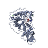

 PDBj
PDBj

 Assembly
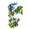


 Mass: 65.409 Da / Num. of mol.: 2 / Source method: obtained synthetically / Formula: Zn
 Mass: 18.015 Da / Num. of mol.: 242 / Source method: isolated from a natural source / Formula: H2O
 Sample preparation
 / Beamline: ID23-1 / Wavelength: 0.984 Å
 Processing