 Movie
Movie Controller

[English] 日本語
 Yorodumi
- PDB-2pk0: Structure of the S. agalactiae serine/threonine phosphatase at 2.... -
+ Open data

- Basic information
| Entry | Database: PDB / ID: 2pk0 | ||||||
|---|---|---|---|---|---|---|---|
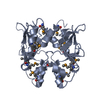
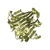
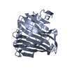
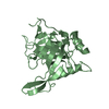
| Title | Structure of the S. agalactiae serine/threonine phosphatase at 2.65 resolution | ||||||
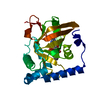
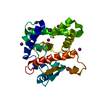
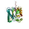
 Components Components | Serine/threonine protein phosphatase Stp1 | ||||||
 Keywords Keywords | SIGNALING PROTEIN / Streptococcus agalactiae / serine / threonine / phosphatase / signaling motif | ||||||
| Function / homology |  Function and homology information Function and homology informationprotein-serine/threonine phosphatase / protein serine/threonine phosphatase activity / phosphatase activity / metal ion binding Similarity search - Function | ||||||
| Biological species |  Streptococcus agalactiae (bacteria) Streptococcus agalactiae (bacteria) | ||||||
| Method |  X-RAY DIFFRACTION / SYNCHROTRON / MOLECULAR REPLACEMENT / Resolution: 2.65 Å X-RAY DIFFRACTION / SYNCHROTRON / MOLECULAR REPLACEMENT / Resolution: 2.65 Å | ||||||
 Authors Authors | Rantanen, M.K. / Lehtio, L. / Rajagopal, L. / Rubens, C.E. / Goldman, A. | ||||||
 Citation Citation | Journal: Febs J. / Year: 2007 Title: Structure of Streptococcus agalactiae serine/threonine phosphatase. The subdomain conformation is coupled to the binding of a third metal ion Authors: Rantanen, M.K. / Lehtio, L. / Rajagopal, L. / Rubens, C.E. / Goldman, A. | ||||||
| History |
|
- Structure visualization
| Structure viewer | Molecule: MolmilJmol/JSmol |
|---|
- Downloads & links
-Download
| PDBx/mmCIF format | 2pk0.cif.gz | 202.6 KB | Display | PDBx/mmCIF format |
|---|---|---|---|---|
| PDB format | pdb2pk0.ent.gz | 161 KB | Display | PDB format |
| PDBx/mmJSON format | 2pk0.json.gz | Tree view | PDBx/mmJSON format | |
| Others |  Other downloads Other downloads |
-Validation report
| Arichive directory | https://data.pdbj.org/pub/pdb/validation_reports/pk/2pk0ftp://data.pdbj.org/pub/pdb/validation_reports/pk/2pk0 | HTTPS FTP |
|---|
-Related structure data
| Similar structure data |
|---|

-Links
 PDBj
PDBj- Assembly
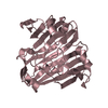
| Deposited unit | 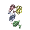
| ||||||||||||||||||||||||||||||||||||||||||||||||||||||||||||||||||||
|---|---|---|---|---|---|---|---|---|---|---|---|---|---|---|---|---|---|---|---|---|---|---|---|---|---|---|---|---|---|---|---|---|---|---|---|---|---|---|---|---|---|---|---|---|---|---|---|---|---|---|---|---|---|---|---|---|---|---|---|---|---|---|---|---|---|---|---|---|---|
| 1 | 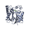
| ||||||||||||||||||||||||||||||||||||||||||||||||||||||||||||||||||||
| 2 | 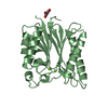
| ||||||||||||||||||||||||||||||||||||||||||||||||||||||||||||||||||||
| 3 | 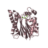
| ||||||||||||||||||||||||||||||||||||||||||||||||||||||||||||||||||||
| 4 | 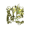
| ||||||||||||||||||||||||||||||||||||||||||||||||||||||||||||||||||||
| Unit cell |
| ||||||||||||||||||||||||||||||||||||||||||||||||||||||||||||||||||||
| Noncrystallographic symmetry (NCS) | NCS domain:
NCS domain segments: Component-ID: 1 / Refine code: 4
NCS ensembles :
| ||||||||||||||||||||||||||||||||||||||||||||||||||||||||||||||||||||
| Details | Biological monomer |
-Components
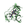
| #1: Protein | Mass: 27404.447 Da / Num. of mol.: 4 Source method: isolated from a genetically manipulated source Source: (gene. exp.) Streptococcus agalactiae (bacteria) / Strain: A909 / Gene: stp / Plasmid: pGEX4T3 / Species (production host): Escherichia coli / Production host: References: UniProt: Q3K363, UniProt: Q8VQA1*PLUS, protein-serine/threonine phosphatase #2: Chemical | ChemComp-MG /   Mass: 24.305 Da / Num. of mol.: 10 / Source method: obtained synthetically / Formula: Mg Mass: 24.305 Da / Num. of mol.: 10 / Source method: obtained synthetically / Formula: Mg#3: Chemical | ChemComp-GOL / |   Mass: 92.094 Da / Num. of mol.: 1 / Source method: obtained synthetically / Formula: C3H8O3 Mass: 92.094 Da / Num. of mol.: 1 / Source method: obtained synthetically / Formula: C3H8O3#4: Chemical | ChemComp-CL / |   Mass: 35.453 Da / Num. of mol.: 1 / Source method: obtained synthetically / Formula: Cl Mass: 35.453 Da / Num. of mol.: 1 / Source method: obtained synthetically / Formula: Cl#5: Water | ChemComp-HOH / |  Mass: 18.015 Da / Num. of mol.: 295 / Source method: isolated from a natural source / Formula: H2O Mass: 18.015 Da / Num. of mol.: 295 / Source method: isolated from a natural source / Formula: H2O |
|---|
-Experimental details
-Experiment
| Experiment | Method: X-RAY DIFFRACTION / Number of used crystals: 1 |
|---|
- Sample preparation
| Crystal | Density Matthews: 2.54 Å3/Da / Density % sol: 51.65 % |
|---|---|
| Crystal grow | Temperature: 298 K / Method: vapor diffusion, hanging drop / pH: 8.5 Details: 0.2 M Mg acetate, 18% PEG 8000, 0.1M Tris, pH 8.5, VAPOR DIFFUSION, HANGING DROP, temperature 298K |
-Data collection
| Diffraction | Mean temperature: 100 K |
|---|---|
| Diffraction source | Source: SYNCHROTRON / Site: ESRF  / Beamline: ID14-1 / Wavelength: 0.933 Å / Beamline: ID14-1 / Wavelength: 0.933 Å |
| Detector | Type: MARRESEARCH / Detector: IMAGE PLATE / Date: May 20, 2005 |
| Radiation | Protocol: SINGLE WAVELENGTH / Monochromatic (M) / Laue (L): M / Scattering type: x-ray |
| Radiation wavelength | Wavelength: 0.933 Å / Relative weight: 1 |
| Reflection | Resolution: 2.65→20 Å / Num. all: 32931 / Num. obs: 32767 / % possible obs: 99.5 % / Observed criterion σ(F): 2 / Observed criterion σ(I): 3 / Redundancy: 5.2 % / Biso Wilson estimate: 50 Å2 / Rmerge(I) obs: 0.115 / Rsym value: 0.115 / Net I/σ(I): 11.7 |
| Reflection shell | Resolution: 2.65→2.7 Å / Redundancy: 4.2 % / Rmerge(I) obs: 0.44 / Mean I/σ(I) obs: 3.7 / Num. unique all: 7442 / Rsym value: 0.44 / % possible all: 99 |
- Processing
| Software |
| |||||||||||||||||||||||||||||||||||||||||||||||||||||||||||||||||||||||||||||||||||||||||||||||
|---|---|---|---|---|---|---|---|---|---|---|---|---|---|---|---|---|---|---|---|---|---|---|---|---|---|---|---|---|---|---|---|---|---|---|---|---|---|---|---|---|---|---|---|---|---|---|---|---|---|---|---|---|---|---|---|---|---|---|---|---|---|---|---|---|---|---|---|---|---|---|---|---|---|---|---|---|---|---|---|---|---|---|---|---|---|---|---|---|---|---|---|---|---|---|---|---|
| Refinement | Method to determine structure: MOLECULAR REPLACEMENT Starting model: Manually built model from SAD phasing = S.a. STP with 70% of residues built Resolution: 2.65→19.65 Å / Cor.coef. Fo:Fc: 0.929 / Cor.coef. Fo:Fc free: 0.87 / SU B: 12.859 / SU ML: 0.273 / Cross valid method: THROUGHOUT / σ(F): 2 / σ(I): 2 / ESU R: 3.077 / ESU R Free: 0.364 / Stereochemistry target values: MAXIMUM LIKELIHOOD / Details: HYDROGENS HAVE BEEN ADDED IN THE RIDING POSITIONS
| |||||||||||||||||||||||||||||||||||||||||||||||||||||||||||||||||||||||||||||||||||||||||||||||
| Solvent computation | Ion probe radii: 0.8 Å / Shrinkage radii: 0.8 Å / VDW probe radii: 1.4 Å / Solvent model: BABINET MODEL WITH MASK | |||||||||||||||||||||||||||||||||||||||||||||||||||||||||||||||||||||||||||||||||||||||||||||||
| Displacement parameters | Biso mean: 42.058 Å2
| |||||||||||||||||||||||||||||||||||||||||||||||||||||||||||||||||||||||||||||||||||||||||||||||
| Refinement step | Cycle: LAST / Resolution: 2.65→19.65 Å
| |||||||||||||||||||||||||||||||||||||||||||||||||||||||||||||||||||||||||||||||||||||||||||||||
| Refine LS restraints |
| |||||||||||||||||||||||||||||||||||||||||||||||||||||||||||||||||||||||||||||||||||||||||||||||
| Refine LS restraints NCS | Dom-ID: 1 / Refine-ID: X-RAY DIFFRACTION
| |||||||||||||||||||||||||||||||||||||||||||||||||||||||||||||||||||||||||||||||||||||||||||||||
| LS refinement shell | Resolution: 2.65→2.718 Å / Total num. of bins used: 20
|
