 Movie
Movie Controller

[English] 日本語
 Yorodumi
- PDB-6ihw: Crystal structure of bacterial serine phosphatase bearing R161K m... -
+ Open data

- Basic information
| Entry | Database: PDB / ID: 6ihw | ||||||
|---|---|---|---|---|---|---|---|
| Title | Crystal structure of bacterial serine phosphatase bearing R161K mutation | ||||||
 Components Components | Phosphorylated protein phosphatase | ||||||
 Keywords Keywords | HYDROLASE / bacteria / phosphatase / metal binding | ||||||
| Function / homology |  Function and homology information Function and homology informationprotein-serine/threonine phosphatase / protein serine/threonine phosphatase activity / metal ion binding Similarity search - Function | ||||||
| Biological species |   Staphylococcus aureus (bacteria) Staphylococcus aureus (bacteria) | ||||||
| Method |  X-RAY DIFFRACTION / SYNCHROTRON / MOLECULAR REPLACEMENT / Resolution: 1.55 Å X-RAY DIFFRACTION / SYNCHROTRON / MOLECULAR REPLACEMENT / Resolution: 1.55 Å | ||||||
 Authors Authors | Yang, C.-G. / yang, T. | ||||||
| Funding support |  China, 1items China, 1items
| ||||||
 Citation Citation | Journal: Acs Infect Dis. / Year: 2019 Title: Structural Insight into the Mechanism of Staphylococcus aureus Stp1 Phosphatase. Authors: Yang, T. / Liu, T. / Gan, J. / Yu, K. / Chen, K. / Xue, W. / Lan, L. / Yang, S. / Yang, C.G. | ||||||
| History |
|
- Structure visualization
| Structure viewer | Molecule: MolmilJmol/JSmol |
|---|
- Downloads & links
-Download
| PDBx/mmCIF format | 6ihw.cif.gz | 123.2 KB | Display | PDBx/mmCIF format |
|---|---|---|---|---|
| PDB format | pdb6ihw.ent.gz | 92.6 KB | Display | PDB format |
| PDBx/mmJSON format | 6ihw.json.gz | Tree view | PDBx/mmJSON format | |
| Others |  Other downloads Other downloads |
-Validation report
| Arichive directory | https://data.pdbj.org/pub/pdb/validation_reports/ih/6ihwftp://data.pdbj.org/pub/pdb/validation_reports/ih/6ihw | HTTPS FTP |
|---|
-Related structure data
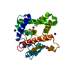
| Related structure data |  6ihlC  6ihrC  6ihsC  6ihtC 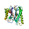 6ihuC 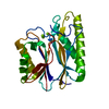 6ihvC 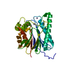 5f1mS S: Starting model for refinement C: citing same article ( |
|---|---|
| Similar structure data |


-Links
 PDBj
PDBj- Assembly
| Deposited unit | 
| ||||||||
|---|---|---|---|---|---|---|---|---|---|
| 1 |
| ||||||||
| Unit cell |
|
-Components
| #1: Protein | Mass: 30109.648 Da / Num. of mol.: 1 / Mutation: R161K Source method: isolated from a genetically manipulated source Source: (gene. exp.) Staphylococcus aureus (bacteria)Gene: prpC, prpC_1, BN1321_240063, BTN44_06615, CSC83_01010, CSC87_08725, EP54_08495, EQ90_08165, ERS072840_01404, NCTC11940_01141, NCTC13131_00423, NCTC13196_02843, NCTC9944_01222, RK64_06500, ...Gene: prpC, prpC_1, BN1321_240063, BTN44_06615, CSC83_01010, CSC87_08725, EP54_08495, EQ90_08165, ERS072840_01404, NCTC11940_01141, NCTC13131_00423, NCTC13196_02843, NCTC9944_01222, RK64_06500, SAMEA1469870_01594, SAMEA1531701_01402 Production host: References: UniProt: Q9RL81, protein-serine/threonine phosphatase | ||
|---|---|---|---|
| #2: Chemical | ChemComp-MG /   Mass: 24.305 Da / Num. of mol.: 4 / Source method: obtained synthetically / Formula: Mg Mass: 24.305 Da / Num. of mol.: 4 / Source method: obtained synthetically / Formula: Mg#3: Water | ChemComp-HOH / |  Mass: 18.015 Da / Num. of mol.: 173 / Source method: isolated from a natural source / Formula: H2O Mass: 18.015 Da / Num. of mol.: 173 / Source method: isolated from a natural source / Formula: H2O |
-Experimental details
-Experiment
| Experiment | Method: X-RAY DIFFRACTION / Number of used crystals: 1 |
|---|
- Sample preparation
| Crystal | Density Matthews: 1.93 Å3/Da / Density % sol: 36.18 % |
|---|---|
| Crystal grow | Temperature: 289 K / Method: vapor diffusion, sitting drop / Details: 0.05 M MgCl2, 0.1 M HEPES (pH=7.5), 30% PEG 3350. |
-Data collection
| Diffraction | Mean temperature: 100 K / Serial crystal experiment: N |
|---|---|
| Diffraction source | Source: SYNCHROTRON / Site: SSRF / Beamline: BL19U1 / Wavelength: 0.9375 Å |
| Detector | Type: DECTRIS PILATUS 6M / Detector: PIXEL / Date: Nov 29, 2017 |
| Radiation | Protocol: SINGLE WAVELENGTH / Monochromatic (M) / Laue (L): M / Scattering type: x-ray |
| Radiation wavelength | Wavelength: 0.9375 Å / Relative weight: 1 |
| Reflection | Resolution: 1.55→30 Å / Num. obs: 32616 / % possible obs: 97.4 % / Redundancy: 4.4 % / Rmerge(I) obs: 0.089 / Net I/σ(I): 12.7 |
| Reflection shell | Resolution: 1.55→1.61 Å / Rmerge(I) obs: 0.198 / Num. unique obs: 3033 |
- Processing
| Software |
| ||||||||||||||||||||||||||||||||||||||||||||||||||||||||||||||||||||||||||||||||||||
|---|---|---|---|---|---|---|---|---|---|---|---|---|---|---|---|---|---|---|---|---|---|---|---|---|---|---|---|---|---|---|---|---|---|---|---|---|---|---|---|---|---|---|---|---|---|---|---|---|---|---|---|---|---|---|---|---|---|---|---|---|---|---|---|---|---|---|---|---|---|---|---|---|---|---|---|---|---|---|---|---|---|---|---|---|---|
| Refinement | Method to determine structure: MOLECULAR REPLACEMENT Starting model: 5F1M Resolution: 1.55→29.606 Å / SU ML: 0.12 / Cross valid method: THROUGHOUT / σ(F): 1.54 / Phase error: 15.98
| ||||||||||||||||||||||||||||||||||||||||||||||||||||||||||||||||||||||||||||||||||||
| Solvent computation | Shrinkage radii: 0.9 Å / VDW probe radii: 1.11 Å | ||||||||||||||||||||||||||||||||||||||||||||||||||||||||||||||||||||||||||||||||||||
| Refinement step | Cycle: LAST / Resolution: 1.55→29.606 Å
| ||||||||||||||||||||||||||||||||||||||||||||||||||||||||||||||||||||||||||||||||||||
| Refine LS restraints |
| ||||||||||||||||||||||||||||||||||||||||||||||||||||||||||||||||||||||||||||||||||||
| LS refinement shell |
| ||||||||||||||||||||||||||||||||||||||||||||||||||||||||||||||||||||||||||||||||||||
| Refinement TLS params. | Method: refined / Origin x: -3.6519 Å / Origin y: 2.8494 Å / Origin z: 79.3242 Å
| ||||||||||||||||||||||||||||||||||||||||||||||||||||||||||||||||||||||||||||||||||||
| Refinement TLS group | Selection details: all |
