 Movie
Movie Controller

[English] 日本語
 Yorodumi
- PDB-2cm1: Crystal structure of the catalytic domain of serine threonine pro... -
+ Open data

- Basic information
| Entry | Database: PDB / ID: 2cm1 | ||||||
|---|---|---|---|---|---|---|---|
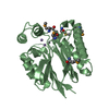
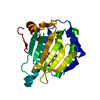
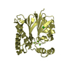
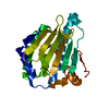
| Title | Crystal structure of the catalytic domain of serine threonine protein phosphatase PstP in complex with 2 Manganese ions. | ||||||
 Components Components | SERINE THREONINE PROTEIN PHOSPHATASE PSTP | ||||||
 Keywords Keywords | HYDROLASE / SER/THR PROTEIN PHOSPHATASE PSTP / HYDROLASE HYPOTHETICAL PROTEIN / MYCOBACTERIUM TUBERCULOSIS | ||||||
| Function / homology |  Function and homology information Function and homology informationprotein-serine/threonine phosphatase / protein serine/threonine phosphatase activity / membrane => GO:0016020 / phosphoprotein phosphatase activity / peptidoglycan-based cell wall / manganese ion binding / regulation of DNA-templated transcription / magnesium ion binding / signal transduction / membrane ...protein-serine/threonine phosphatase / protein serine/threonine phosphatase activity / membrane => GO:0016020 / phosphoprotein phosphatase activity / peptidoglycan-based cell wall / manganese ion binding / regulation of DNA-templated transcription / magnesium ion binding / signal transduction / membrane / metal ion binding / plasma membrane Similarity search - Function | ||||||
| Biological species |   MYCOBACTERIUM TUBERCULOSIS (bacteria) MYCOBACTERIUM TUBERCULOSIS (bacteria) | ||||||
| Method |  X-RAY DIFFRACTION / SYNCHROTRON / MOLECULAR REPLACEMENT / Resolution: 2 Å X-RAY DIFFRACTION / SYNCHROTRON / MOLECULAR REPLACEMENT / Resolution: 2 Å | ||||||
 Authors Authors | Wehenkel, A. / Villarino, A. / Bellinzoni, M. / Alzari, P.M. | ||||||
 Citation Citation | Journal: J.Mol.Biol. / Year: 2007 Title: Structural and Binding Studies of the Three-Metal Center in Two Mycobacterial Ppm Ser/Thr Protein Phosphatases. Authors: Wehenkel, A. / Bellinzoni, M. / Schaeffer, F. / Villarino, A. / Alzari, P.M. | ||||||
| History |
| ||||||
| Remark 700 | SHEET THE SHEET STRUCTURE OF THIS MOLECULE IS BIFURCATED. IN ORDER TO REPRESENT THIS FEATURE IN ... SHEET THE SHEET STRUCTURE OF THIS MOLECULE IS BIFURCATED. IN ORDER TO REPRESENT THIS FEATURE IN THE SHEET RECORDS BELOW, TWO SHEETS ARE DEFINED. |
- Structure visualization
| Structure viewer | Molecule: MolmilJmol/JSmol |
|---|
- Downloads & links
-Download
| PDBx/mmCIF format | 2cm1.cif.gz | 61.2 KB | Display | PDBx/mmCIF format |
|---|---|---|---|---|
| PDB format | pdb2cm1.ent.gz | 43.1 KB | Display | PDB format |
| PDBx/mmJSON format | 2cm1.json.gz | Tree view | PDBx/mmJSON format | |
| Others |  Other downloads Other downloads |
-Validation report
| Arichive directory | https://data.pdbj.org/pub/pdb/validation_reports/cm/2cm1ftp://data.pdbj.org/pub/pdb/validation_reports/cm/2cm1 | HTTPS FTP |
|---|
-Related structure data
| Related structure data |  2v06C  1txoS S: Starting model for refinement C: citing same article ( |
|---|---|
| Similar structure data |


-Links
 PDBj
PDBj- Assembly
| Deposited unit | 
| ||||||||
|---|---|---|---|---|---|---|---|---|---|
| 1 |
| ||||||||
| Unit cell |
|
-Components
| #1: Protein | Mass: 27633.984 Da / Num. of mol.: 1 / Fragment: CATALYTIC DOMAIN, RESIDUES 1-240 Source method: isolated from a genetically manipulated source Details: 2 MANGANESE IONS BOUND IN THE ACTIVE SITE / Source: (gene. exp.) MYCOBACTERIUM TUBERCULOSIS (bacteria) / Strain: H37RV / Plasmid: PET28A / Production host: References: UniProt: P71588, UniProt: P9WHW5*PLUS, protein-serine/threonine phosphatase | ||||
|---|---|---|---|---|---|
| #2: Chemical |   Mass: 54.938 Da / Num. of mol.: 2 / Source method: obtained synthetically / Formula: Mn Mass: 54.938 Da / Num. of mol.: 2 / Source method: obtained synthetically / Formula: Mn#3: Chemical | ChemComp-GOL / |   Mass: 92.094 Da / Num. of mol.: 1 / Source method: obtained synthetically / Formula: C3H8O3 Mass: 92.094 Da / Num. of mol.: 1 / Source method: obtained synthetically / Formula: C3H8O3#4: Water | ChemComp-HOH / |  Mass: 18.015 Da / Num. of mol.: 143 / Source method: isolated from a natural source / Formula: H2O Mass: 18.015 Da / Num. of mol.: 143 / Source method: isolated from a natural source / Formula: H2O |
-Experimental details
-Experiment
| Experiment | Method: X-RAY DIFFRACTION |
|---|
- Sample preparation
| Crystal | Density Matthews: 2.2 Å3/Da / Density % sol: 44 % Description: REJECTION CRITERIA FOR RESOLUTION CUTOFF AT MERGING R VALUE BELOW 0.4 |
|---|---|
| Crystal grow | Details: 14% PEG 8000, 50MM KH2PO4 PH6.5, 15% GLYCEROL |
-Data collection
| Diffraction | Mean temperature: 100 K |
|---|---|
| Diffraction source | Source: SYNCHROTRON / Site: ESRF  / Beamline: ID14-2 / Wavelength: 0.933 / Beamline: ID14-2 / Wavelength: 0.933 |
| Detector | Type: ADSC CCD / Detector: CCD / Date: Feb 15, 2005 / Details: MIRRORS |
| Radiation | Monochromator: DIAMOND (111), GE(220) / Protocol: SINGLE WAVELENGTH / Monochromatic (M) / Laue (L): M / Scattering type: x-ray |
| Radiation wavelength | Wavelength: 0.933 Å / Relative weight: 1 |
| Reflection | Resolution: 2→60 Å / Num. obs: 15969 / % possible obs: 92.8 % / Observed criterion σ(I): 3.96 / Redundancy: 3.5 % / Rmerge(I) obs: 0.09 / Net I/σ(I): 11.67 |
| Reflection shell | Resolution: 2→2.2 Å / Redundancy: 3.5 % / Rmerge(I) obs: 0.4 / Mean I/σ(I) obs: 3.96 / % possible all: 84.1 |
- Processing
| Software |
| ||||||||||||||||||||||||||||||||||||||||||||||||||||||||||||||||||||||||||||||||||||||||||||||||||||||||||||||||||||||||||||||||||||||||||||||||||||||||||||||||||||||||||||||||||||||
|---|---|---|---|---|---|---|---|---|---|---|---|---|---|---|---|---|---|---|---|---|---|---|---|---|---|---|---|---|---|---|---|---|---|---|---|---|---|---|---|---|---|---|---|---|---|---|---|---|---|---|---|---|---|---|---|---|---|---|---|---|---|---|---|---|---|---|---|---|---|---|---|---|---|---|---|---|---|---|---|---|---|---|---|---|---|---|---|---|---|---|---|---|---|---|---|---|---|---|---|---|---|---|---|---|---|---|---|---|---|---|---|---|---|---|---|---|---|---|---|---|---|---|---|---|---|---|---|---|---|---|---|---|---|---|---|---|---|---|---|---|---|---|---|---|---|---|---|---|---|---|---|---|---|---|---|---|---|---|---|---|---|---|---|---|---|---|---|---|---|---|---|---|---|---|---|---|---|---|---|---|---|---|---|
| Refinement | Method to determine structure: MOLECULAR REPLACEMENT Starting model: PDB ENTRY 1TXO Resolution: 2→59.87 Å / Cor.coef. Fo:Fc: 0.946 / Cor.coef. Fo:Fc free: 0.933 / SU B: 4.674 / SU ML: 0.128 / Cross valid method: THROUGHOUT / ESU R: 0.198 / ESU R Free: 0.176 / Stereochemistry target values: MAXIMUM LIKELIHOOD / Details: HYDROGENS HAVE BEEN ADDED IN THE RIDING POSITIONS.
| ||||||||||||||||||||||||||||||||||||||||||||||||||||||||||||||||||||||||||||||||||||||||||||||||||||||||||||||||||||||||||||||||||||||||||||||||||||||||||||||||||||||||||||||||||||||
| Solvent computation | Ion probe radii: 0.8 Å / Shrinkage radii: 0.8 Å / VDW probe radii: 1.2 Å / Solvent model: MASK | ||||||||||||||||||||||||||||||||||||||||||||||||||||||||||||||||||||||||||||||||||||||||||||||||||||||||||||||||||||||||||||||||||||||||||||||||||||||||||||||||||||||||||||||||||||||
| Displacement parameters | Biso mean: 25.87 Å2
| ||||||||||||||||||||||||||||||||||||||||||||||||||||||||||||||||||||||||||||||||||||||||||||||||||||||||||||||||||||||||||||||||||||||||||||||||||||||||||||||||||||||||||||||||||||||
| Refinement step | Cycle: LAST / Resolution: 2→59.87 Å
| ||||||||||||||||||||||||||||||||||||||||||||||||||||||||||||||||||||||||||||||||||||||||||||||||||||||||||||||||||||||||||||||||||||||||||||||||||||||||||||||||||||||||||||||||||||||
| Refine LS restraints |
|