 Movie
Movie Controller

[English] 日本語
 Yorodumi
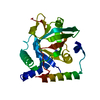
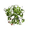
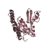
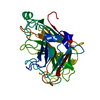
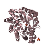
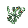
- PDB-4xzj: Crystal structure of ADP-ribosyltransferase Vis in complex with NAD -
+ Open data

- Basic information
| Entry | Database: PDB / ID: 4xzj | ||||||
|---|---|---|---|---|---|---|---|
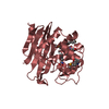
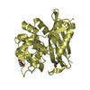
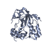
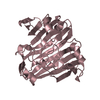
| Title | Crystal structure of ADP-ribosyltransferase Vis in complex with NAD | ||||||
 Components Components | Putative NAD(+)--arginine ADP-ribosyltransferase Vis | ||||||
 Keywords Keywords | TRANSFERASE | ||||||
| Function / homology |  Function and homology information Function and homology informationNAD+-protein-arginine ADP-ribosyltransferase / NAD+-protein-arginine ADP-ribosyltransferase activity / nucleotidyltransferase activity / toxin activity / nucleotide binding / extracellular region Similarity search - Function | ||||||
| Biological species |  Vibrio splendidus (bacteria) Vibrio splendidus (bacteria) | ||||||
| Method |  X-RAY DIFFRACTION / MOLECULAR REPLACEMENT / Resolution: 1.8 Å X-RAY DIFFRACTION / MOLECULAR REPLACEMENT / Resolution: 1.8 Å | ||||||
 Authors Authors | Pfoh, R. / Ravulapalli, R. / Merrill, A.R. / Pai, E.F. | ||||||
 Citation Citation | Journal: Biochemistry / Year: 2015 Title: Characterization of Vis Toxin, a Novel ADP-Ribosyltransferase from Vibrio splendidus. Authors: Ravulapalli, R. / Lugo, M.R. / Pfoh, R. / Visschedyk, D. / Poole, A. / Fieldhouse, R.J. / Pai, E.F. / Merrill, A.R. | ||||||
| History |
|
- Structure visualization
| Structure viewer | Molecule: MolmilJmol/JSmol |
|---|
- Downloads & links
-Download
| PDBx/mmCIF format | 4xzj.cif.gz | 107.8 KB | Display | PDBx/mmCIF format |
|---|---|---|---|---|
| PDB format | pdb4xzj.ent.gz | 81.4 KB | Display | PDB format |
| PDBx/mmJSON format | 4xzj.json.gz | Tree view | PDBx/mmJSON format | |
| Others |  Other downloads Other downloads |
-Validation report
| Arichive directory | https://data.pdbj.org/pub/pdb/validation_reports/xz/4xzjftp://data.pdbj.org/pub/pdb/validation_reports/xz/4xzj | HTTPS FTP |
|---|
-Related structure data
| Related structure data | 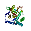 4y1wSC 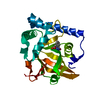 4yc0C S: Starting model for refinement C: citing same article ( |
|---|---|
| Similar structure data |

-Links
 PDBj
PDBj- Assembly
| Deposited unit | 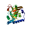
| ||||||||
|---|---|---|---|---|---|---|---|---|---|
| 1 |
| ||||||||
| Unit cell |
| ||||||||
| Details | biological unit is the same as asym. |
-Components
| #1: Protein | Mass: 27037.045 Da / Num. of mol.: 1 / Fragment: residues 20-240 Source method: isolated from a genetically manipulated source Source: (gene. exp.) Vibrio splendidus (bacteria) / Gene: V12B01_18061 / Plasmid: pet 28-MHL / Production host: References: UniProt: A3UNN4, NAD+-protein-arginine ADP-ribosyltransferase |
|---|---|
| #2: Chemical | ChemComp-NAD /   Mass: 663.425 Da / Num. of mol.: 1 / Source method: obtained synthetically / Formula: C21H27N7O14P2 / Comment: NAD*YM Mass: 663.425 Da / Num. of mol.: 1 / Source method: obtained synthetically / Formula: C21H27N7O14P2 / Comment: NAD*YM |
| #3: Water | ChemComp-HOH /  Mass: 18.015 Da / Num. of mol.: 145 / Source method: isolated from a natural source / Formula: H2O Mass: 18.015 Da / Num. of mol.: 145 / Source method: isolated from a natural source / Formula: H2O |
-Experimental details
-Experiment
| Experiment | Method: X-RAY DIFFRACTION / Number of used crystals: 1 |
|---|
- Sample preparation
| Crystal | Density Matthews: 2.61 Å3/Da / Density % sol: 52.8 % |
|---|---|
| Crystal grow | Temperature: 298 K / Method: vapor diffusion, hanging drop / pH: 7 / Details: 30% Jeffamine ED-2001, 0.1 M Hepes |
-Data collection
| Diffraction | Mean temperature: 100 K |
|---|---|
| Diffraction source | Source: ROTATING ANODE / Type: RIGAKU MICROMAX-007 HF / Wavelength: 1.54178 Å |
| Detector | Type: MAR scanner 345 mm plate / Detector: IMAGE PLATE / Date: Aug 29, 2011 |
| Radiation | Monochromator: multi-layer / Protocol: SINGLE WAVELENGTH / Monochromatic (M) / Laue (L): M / Scattering type: x-ray |
| Radiation wavelength | Wavelength: 1.54178 Å / Relative weight: 1 |
| Reflection | Resolution: 1.8→20 Å / Num. obs: 23562 / % possible obs: 94.7 % / Redundancy: 6.25 % / Rmerge(I) obs: 0.0445 / Net I/σ(I): 25.84 |
| Reflection shell | Resolution: 1.8→1.9 Å / Redundancy: 5.24 % / Rmerge(I) obs: 0.1799 / Mean I/σ(I) obs: 8.67 / % possible all: 91.8 |
- Processing
| Software |
| |||||||||||||||||||||||||||||||||||||||||||||||||||||||||||||||||||||||||||
|---|---|---|---|---|---|---|---|---|---|---|---|---|---|---|---|---|---|---|---|---|---|---|---|---|---|---|---|---|---|---|---|---|---|---|---|---|---|---|---|---|---|---|---|---|---|---|---|---|---|---|---|---|---|---|---|---|---|---|---|---|---|---|---|---|---|---|---|---|---|---|---|---|---|---|---|---|
| Refinement | Method to determine structure: MOLECULAR REPLACEMENT Starting model: 4Y1W Resolution: 1.8→18.63 Å / Cor.coef. Fo:Fc: 0.959 / Cor.coef. Fo:Fc free: 0.946 / WRfactor Rfree: 0.2105 / WRfactor Rwork: 0.1802 / FOM work R set: 0.8824 / SU B: 4.226 / SU ML: 0.069 / SU R Cruickshank DPI: 0.1222 / SU Rfree: 0.1147 / Cross valid method: THROUGHOUT / σ(F): 0 / ESU R: 0.122 / ESU R Free: 0.115 / Stereochemistry target values: MAXIMUM LIKELIHOOD Details: HYDROGENS HAVE BEEN ADDED IN THE RIDING POSITIONS U VALUES : WITH TLS ADDED
| |||||||||||||||||||||||||||||||||||||||||||||||||||||||||||||||||||||||||||
| Solvent computation | Ion probe radii: 0.8 Å / Shrinkage radii: 0.8 Å / VDW probe radii: 1.2 Å / Solvent model: BABINET MODEL WITH MASK | |||||||||||||||||||||||||||||||||||||||||||||||||||||||||||||||||||||||||||
| Displacement parameters | Biso max: 66.56 Å2 / Biso mean: 23.386 Å2 / Biso min: 13.86 Å2
| |||||||||||||||||||||||||||||||||||||||||||||||||||||||||||||||||||||||||||
| Refinement step | Cycle: final / Resolution: 1.8→18.63 Å
| |||||||||||||||||||||||||||||||||||||||||||||||||||||||||||||||||||||||||||
| Refine LS restraints |
| |||||||||||||||||||||||||||||||||||||||||||||||||||||||||||||||||||||||||||
| LS refinement shell | Resolution: 1.8→1.846 Å / Total num. of bins used: 20
| |||||||||||||||||||||||||||||||||||||||||||||||||||||||||||||||||||||||||||
| Refinement TLS params. | Method: refined / Refine-ID: X-RAY DIFFRACTION
| |||||||||||||||||||||||||||||||||||||||||||||||||||||||||||||||||||||||||||
| Refinement TLS group |
|
