 Movie
Movie Controller

[English] 日本語
 Yorodumi
- PDB-7kym: Crystal structure of the MarR family transcriptional regulator fr... -
+ Open data

- Basic information
| Entry | Database: PDB / ID: 7kym | ||||||
|---|---|---|---|---|---|---|---|
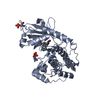
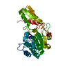
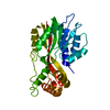
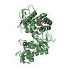
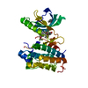
| Title | Crystal structure of the MarR family transcriptional regulator from Bradyrhizobium japonicum | ||||||
 Components Components | MarR family transcriptional regulator | ||||||
 Keywords Keywords | DNA BINDING PROTEIN / Transcriptional Regulator / Ligand Binding | ||||||
| Function / homology |  Function and homology information Function and homology information | ||||||
| Biological species |  Bradyrhizobium japonicum (bacteria) Bradyrhizobium japonicum (bacteria) | ||||||
| Method |  X-RAY DIFFRACTION / SYNCHROTRON / MOLECULAR REPLACEMENT / Resolution: 1.5 Å X-RAY DIFFRACTION / SYNCHROTRON / MOLECULAR REPLACEMENT / Resolution: 1.5 Å | ||||||
 Authors Authors | Walton, W.G. / Redinbo, M.R. / Dangl, J.L. | ||||||
| Funding support |  United States, 1items United States, 1items
| ||||||
 Citation Citation | Journal: Nat Microbiol / Year: 2022 Title: Diverse MarR bacterial regulators of auxin catabolism in the plant microbiome. Authors: Conway, J.M. / Walton, W.G. / Salas-Gonzalez, I. / Law, T.F. / Lindberg, C.A. / Crook, L.E. / Kosina, S.M. / Fitzpatrick, C.R. / Lietzan, A.D. / Northen, T.R. / Jones, C.D. / Finkel, O.M. / ...Authors: Conway, J.M. / Walton, W.G. / Salas-Gonzalez, I. / Law, T.F. / Lindberg, C.A. / Crook, L.E. / Kosina, S.M. / Fitzpatrick, C.R. / Lietzan, A.D. / Northen, T.R. / Jones, C.D. / Finkel, O.M. / Redinbo, M.R. / Dangl, J.L. | ||||||
| History |
|
- Structure visualization
| Structure viewer | Molecule: MolmilJmol/JSmol |
|---|
- Downloads & links
-Download
| PDBx/mmCIF format | 7kym.cif.gz | 83 KB | Display | PDBx/mmCIF format |
|---|---|---|---|---|
| PDB format | pdb7kym.ent.gz | 59.3 KB | Display | PDB format |
| PDBx/mmJSON format | 7kym.json.gz | Tree view | PDBx/mmJSON format | |
| Others |  Other downloads Other downloads |
-Validation report
| Arichive directory | https://data.pdbj.org/pub/pdb/validation_reports/ky/7kymftp://data.pdbj.org/pub/pdb/validation_reports/ky/7kym | HTTPS FTP |
|---|
-Related structure data
| Related structure data |  7kfoC  7kfqC 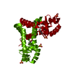 7kfsC  7kh3C  7kigC  7kjlC  7kjqC  7kk0C 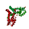 7kkcC 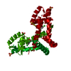 7kkiC  7krhC  7kuaC  7l19C  7l1iC 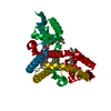 3cjnS S: Starting model for refinement C: citing same article ( |
|---|---|
| Similar structure data |


-Links
 PDBj
PDBj- Assembly
| Deposited unit | 
| ||||||||||||
|---|---|---|---|---|---|---|---|---|---|---|---|---|---|
| 1 | 
| ||||||||||||
| Unit cell |
|
-Components
| #1: Protein | Mass: 21214.062 Da / Num. of mol.: 2 Source method: isolated from a genetically manipulated source Source: (gene. exp.) Bradyrhizobium japonicum (bacteria) / Gene: BJA01nite_12290, MA20_13490 / Production host: #2: Chemical |   Mass: 94.971 Da / Num. of mol.: 2 / Source method: obtained synthetically / Formula: PO4 Mass: 94.971 Da / Num. of mol.: 2 / Source method: obtained synthetically / Formula: PO4#3: Water | ChemComp-HOH / |  Mass: 18.015 Da / Num. of mol.: 293 / Source method: isolated from a natural source / Formula: H2O Mass: 18.015 Da / Num. of mol.: 293 / Source method: isolated from a natural source / Formula: H2OHas ligand of interest | N | |
|---|
-Experimental details
-Experiment
| Experiment | Method: X-RAY DIFFRACTION / Number of used crystals: 1 |
|---|
- Sample preparation
| Crystal grow | Temperature: 293 K / Method: vapor diffusion, sitting drop / pH: 6.5 Details: 0.1 M Bis-Tris: HCl, pH 6.5, 20 % (w/v) PEG MME 5000 |
|---|
-Data collection
| Diffraction | Mean temperature: 100 K / Serial crystal experiment: N |
|---|---|
| Diffraction source | Source: SYNCHROTRON / Site: APS / Beamline: 23-ID-D / Wavelength: 1.0332 Å |
| Detector | Type: DECTRIS PILATUS3 6M / Detector: PIXEL / Date: Dec 5, 2020 |
| Radiation | Protocol: SINGLE WAVELENGTH / Monochromatic (M) / Laue (L): M / Scattering type: x-ray |
| Radiation wavelength | Wavelength: 1.0332 Å / Relative weight: 1 |
| Reflection | Resolution: 1.5→47.26 Å / Num. obs: 44118 / % possible obs: 99.07 % / Redundancy: 2 % / CC1/2: 1 / Net I/σ(I): 16.2 |
| Reflection shell | Resolution: 1.5→1.554 Å / Num. unique obs: 4182 / CC1/2: 0.839 |
- Processing
| Software |
| ||||||||||||||||||||||||
|---|---|---|---|---|---|---|---|---|---|---|---|---|---|---|---|---|---|---|---|---|---|---|---|---|---|
| Refinement | Method to determine structure: MOLECULAR REPLACEMENT Starting model: 3CJN Resolution: 1.5→47.26 Å / Cross valid method: FREE R-VALUE Stereochemistry target values: GeoStd + Monomer Library + CDL v1.2
| ||||||||||||||||||||||||
| Displacement parameters | Biso mean: 24.25 Å2 | ||||||||||||||||||||||||
| Refinement step | Cycle: LAST / Resolution: 1.5→47.26 Å
| ||||||||||||||||||||||||
| Refine LS restraints |
|
