| Entry | Database: PDB / ID: 3k6x
|
|---|
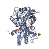
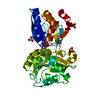
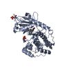
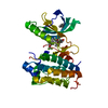
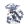
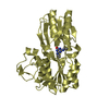
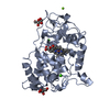
| Title | M. acetivorans Molybdate-Binding Protein (ModA) in Molybdate-Bound Close Form with 2 Molecules in Asymmetric Unit Forming Beta Barrel |
|---|
 Components Components | Solute-binding protein MA_0280 |
|---|
 Keywords Keywords | TRANSPORT PROTEIN / ModA / molybdate / Methanosarcina acetivorans / periplasmic binding protein / ABC transporter / ligand / metal-binding protein |
|---|
| Function / homology |  Function and homology information Function and homology information
Tungstate ABC transporter, substrate-binding protein WtpA / : / Bacterial extracellular solute-binding protein / Periplasmic binding protein-like II / D-Maltodextrin-Binding Protein; domain 2 / 3-Layer(aba) Sandwich / Alpha BetaSimilarity search - Domain/homology |
|---|
| Biological species |  Methanosarcina acetivorans (archaea) Methanosarcina acetivorans (archaea) |
|---|
| Method |  X-RAY DIFFRACTION / SYNCHROTRON / MOLECULAR REPLACEMENT / molecular replacement / Resolution: 2.25 Å X-RAY DIFFRACTION / SYNCHROTRON / MOLECULAR REPLACEMENT / molecular replacement / Resolution: 2.25 Å |
|---|
 Authors Authors | Chan, S. / Chernishof, I. / Giuroiu, I. / Sawaya, M.R. / Chiang, J. / Gunsalus, R.P. / Arbing, M.A. / Perry, L.J. |
|---|
 Citation Citation | Journal: Acta Crystallogr.,Sect.F / Year: 2010
Title: Apo and ligand-bound structures of ModA from the archaeon Methanosarcina acetivorans
Authors: Chan, S. / Giuroiu, I. / Chernishof, I. / Sawaya, M.R. / Chiang, J. / Gunsalus, R.P. / Arbing, M.A. / Perry, L.J. |
|---|
| History | | Deposition | Oct 9, 2009 | Deposition site: RCSB / Processing site: RCSB |
|---|
| Revision 1.0 | Jan 12, 2010 | Provider: repository / Type: Initial release |
|---|
| Revision 1.1 | Jul 13, 2011 | Group: Advisory / Refinement description / Version format compliance |
|---|
| Revision 1.2 | Nov 1, 2017 | Group: Refinement description / Category: software |
|---|
| Revision 1.3 | Feb 21, 2024 | Group: Data collection / Database references ...Data collection / Database references / Derived calculations / Refinement description
Category: chem_comp_atom / chem_comp_bond ...chem_comp_atom / chem_comp_bond / database_2 / struct_ncs_dom_lim / struct_ref_seq_dif / struct_site
Item: _database_2.pdbx_DOI / _database_2.pdbx_database_accession ..._database_2.pdbx_DOI / _database_2.pdbx_database_accession / _struct_ncs_dom_lim.beg_auth_comp_id / _struct_ncs_dom_lim.beg_label_asym_id / _struct_ncs_dom_lim.beg_label_comp_id / _struct_ncs_dom_lim.beg_label_seq_id / _struct_ncs_dom_lim.end_auth_comp_id / _struct_ncs_dom_lim.end_label_asym_id / _struct_ncs_dom_lim.end_label_comp_id / _struct_ncs_dom_lim.end_label_seq_id / _struct_ref_seq_dif.details / _struct_site.pdbx_auth_asym_id / _struct_site.pdbx_auth_comp_id / _struct_site.pdbx_auth_seq_id |
|---|
|
|---|
 Movie
Movie Controller

 Yorodumi
 Open data
 Basic information
 Structure visualization
 Downloads & links
 Other downloads


 PDBj
PDBj Assembly