 Movie
Movie Controller

[English] 日本語
 Yorodumi
- PDB-3k6v: M. acetivorans Molybdate-Binding Protein (ModA) in Citrate-Bound ... -
+ Open data

- Basic information
| Entry | Database: PDB / ID: 3k6v | ||||||
|---|---|---|---|---|---|---|---|
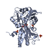
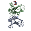
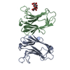
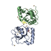
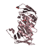
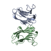
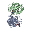
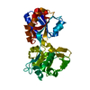
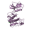
| Title | M. acetivorans Molybdate-Binding Protein (ModA) in Citrate-Bound Open Form | ||||||
 Components Components | Solute-binding protein MA_0280 | ||||||
 Keywords Keywords | TRANSPORT PROTEIN / ModA / molybdate / Methanosarcina acetivorans / periplasmic binding protein / ABC transporter / ligand / metal-binding protein | ||||||
| Function / homology |  Function and homology information Function and homology information | ||||||
| Biological species |  Methanosarcina acetivorans (archaea) Methanosarcina acetivorans (archaea) | ||||||
| Method |  X-RAY DIFFRACTION / SYNCHROTRON / MOLECULAR REPLACEMENT / molecular replacement / Resolution: 1.69 Å X-RAY DIFFRACTION / SYNCHROTRON / MOLECULAR REPLACEMENT / molecular replacement / Resolution: 1.69 Å | ||||||
 Authors Authors | Chan, S. / Giuroiu, I. / Chernishof, I. / Sawaya, M.R. / Chiang, J. / Gunsalus, R.P. / Arbing, M.A. / Perry, L.J. | ||||||
 Citation Citation | Journal: Acta Crystallogr.,Sect.F / Year: 2010 Title: Apo and ligand-bound structures of ModA from the archaeon Methanosarcina acetivorans Authors: Chan, S. / Giuroiu, I. / Chernishof, I. / Sawaya, M.R. / Chiang, J. / Gunsalus, R.P. / Arbing, M.A. / Perry, L.J. | ||||||
| History |
|
- Structure visualization
| Structure viewer | Molecule: MolmilJmol/JSmol |
|---|
- Downloads & links
-Download
| PDBx/mmCIF format | 3k6v.cif.gz | 81.5 KB | Display | PDBx/mmCIF format |
|---|---|---|---|---|
| PDB format | pdb3k6v.ent.gz | 60.2 KB | Display | PDB format |
| PDBx/mmJSON format | 3k6v.json.gz | Tree view | PDBx/mmJSON format | |
| Others |  Other downloads Other downloads |
-Validation report
| Arichive directory | https://data.pdbj.org/pub/pdb/validation_reports/k6/3k6vftp://data.pdbj.org/pub/pdb/validation_reports/k6/3k6v | HTTPS FTP |
|---|
-Related structure data


-Links
 PDBj
PDBj- Assembly
| Deposited unit | 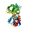
| ||||||||
|---|---|---|---|---|---|---|---|---|---|
| 1 |
| ||||||||
| Unit cell |
|
-Components
| #1: Protein | Mass: 39160.156 Da / Num. of mol.: 1 Source method: isolated from a genetically manipulated source Details: N-terminal TEV-cleavable 6xHis-tag / Source: (gene. exp.) Methanosarcina acetivorans (archaea) / Strain: C2A / Gene: MA0280, MA_0280 / Plasmid: pETM-11 / Production host:  |
|---|---|
| #2: Chemical | ChemComp-CIT /   Mass: 192.124 Da / Num. of mol.: 1 / Source method: obtained synthetically / Formula: C6H8O7 Mass: 192.124 Da / Num. of mol.: 1 / Source method: obtained synthetically / Formula: C6H8O7 |
| #3: Water | ChemComp-HOH /  Mass: 18.015 Da / Num. of mol.: 294 / Source method: isolated from a natural source / Formula: H2O Mass: 18.015 Da / Num. of mol.: 294 / Source method: isolated from a natural source / Formula: H2O |
-Experimental details
-Experiment
| Experiment | Method: X-RAY DIFFRACTION / Number of used crystals: 1 |
|---|
- Sample preparation
| Crystal | Density Matthews: 2.57 Å3/Da / Density % sol: 52.14 % |
|---|---|
| Crystal grow | Temperature: 293 K / Method: vapor diffusion, sitting drop / pH: 6 Details: 4mM Sodium Sulfate, 1.4M Ammonium Citrate, pH6.0, vapor diffusion, sitting drop, temperature 293K |
-Data collection
| Diffraction | Mean temperature: 100 K |
|---|---|
| Diffraction source | Source: SYNCHROTRON / Site: ALS  / Beamline: 8.2.2 / Wavelength: 0.98 Å / Beamline: 8.2.2 / Wavelength: 0.98 Å |
| Detector | Type: ADSC QUANTUM 315 / Detector: CCD / Date: Sep 30, 2005 |
| Radiation | Protocol: SINGLE WAVELENGTH / Scattering type: x-ray |
| Radiation wavelength | Wavelength: 0.98 Å / Relative weight: 1 |
| Reflection | Resolution: 1.69→58.62 Å / Num. obs: 84835 / % possible obs: 96.9 % / Redundancy: 5.8 % / Rmerge(I) obs: 0.054 / Rsym value: 0.04 / Χ2: 0.995 / Net I/σ(I): 16.3 |
| Reflection shell | Resolution: 1.69→1.75 Å / Redundancy: 3.8 % / Rmerge(I) obs: 0.593 / Mean I/σ(I) obs: 2.26 / Num. unique all: 7182 / Rsym value: 0.472 / Χ2: 1.004 / % possible all: 82.3 |
-Phasing
| Phasing | Method: molecular replacement | ||||||
|---|---|---|---|---|---|---|---|
| Phasing MR | Rfactor: 0.29 / Cor.coef. Fo:Fc: 0.76
|
- Processing
| Software |
| |||||||||||||||||||||||||||||||||||||||||||||||||||||||||||||||||
|---|---|---|---|---|---|---|---|---|---|---|---|---|---|---|---|---|---|---|---|---|---|---|---|---|---|---|---|---|---|---|---|---|---|---|---|---|---|---|---|---|---|---|---|---|---|---|---|---|---|---|---|---|---|---|---|---|---|---|---|---|---|---|---|---|---|---|
| Refinement | Method to determine structure: MOLECULAR REPLACEMENT / Resolution: 1.69→58.62 Å / Cor.coef. Fo:Fc: 0.964 / Cor.coef. Fo:Fc free: 0.955 / WRfactor Rfree: 0.217 / WRfactor Rwork: 0.195 / Occupancy max: 1 / Occupancy min: 0 / FOM work R set: 0.865 / SU B: 4.278 / SU ML: 0.065 / SU R Cruickshank DPI: 0.097 / SU Rfree: 0.093 / TLS residual ADP flag: LIKELY RESIDUAL / Cross valid method: THROUGHOUT / σ(F): 0 / ESU R: 0.097 / ESU R Free: 0.093 / Stereochemistry target values: MAXIMUM LIKELIHOOD Details: HYDROGENS HAVE BEEN ADDED IN THE RIDING POSITIONS U VALUES : RESIDUAL ONLY
| |||||||||||||||||||||||||||||||||||||||||||||||||||||||||||||||||
| Solvent computation | Ion probe radii: 0.8 Å / Shrinkage radii: 0.8 Å / VDW probe radii: 1.4 Å / Solvent model: MASK | |||||||||||||||||||||||||||||||||||||||||||||||||||||||||||||||||
| Displacement parameters | Biso max: 65.29 Å2 / Biso mean: 19.883 Å2 / Biso min: 11.15 Å2
| |||||||||||||||||||||||||||||||||||||||||||||||||||||||||||||||||
| Refinement step | Cycle: LAST / Resolution: 1.69→58.62 Å
| |||||||||||||||||||||||||||||||||||||||||||||||||||||||||||||||||
| Refine LS restraints |
| |||||||||||||||||||||||||||||||||||||||||||||||||||||||||||||||||
| LS refinement shell | Resolution: 1.69→1.734 Å / Total num. of bins used: 20
| |||||||||||||||||||||||||||||||||||||||||||||||||||||||||||||||||
| Refinement TLS params. | Method: refined / Origin x: 26.3992 Å / Origin y: 44.2635 Å / Origin z: 14.6913 Å
|
