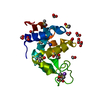
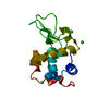
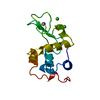
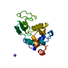
Entry Database : PDB / ID : 5azgTitle Crystal structure of LGG-1 complexed with a UNC-51 peptide Protein lgg-1 Serine/threonine-protein kinase unc-51 Keywords / / Function / homology Function Domain/homology Component
/ / / / / / / / / / / / / / / / / / / / / / / / / / / / / / / / / / / / / / / / / / / / / / / / / / / / / / / / / / / / / / / / / / / / / / / / / / / / / / / / / / / / / / / / / / / / / / / / / / / / / / / / / / / / / / / / / / / / / Biological species Caenorhabditis elegans (invertebrata)Method / / / Resolution : 1.81 Å Authors Watanabe, Y. / Fujioka, Y. / Noda, N.N. Funding support Organization Grant number Country Ministry of Education, Culture, Sports, Science and Technology of Japan 25111004 Japan Science and Technology Agency CREST
Journal : Mol.Cell / Year : 2015Title : Structural Basis of the Differential Function of the Two C. elegans Atg8 Homologs, LGG-1 and LGG-2, in Autophagy.Authors : Wu, F. / Watanabe, Y. / Guo, X.Y. / Qi, X. / Wang, P. / Zhao, H.Y. / Wang, Z. / Fujioka, Y. / Zhang, H. / Ren, J.Q. / Fang, T.C. / Shen, Y.X. / Feng, W. / Hu, J.J. / Noda, N.N. / Zhang, H. History Deposition Oct 5, 2015 Deposition site / Processing site Revision 1.0 Dec 30, 2015 Provider / Type Revision 1.1 Jan 1, 2020 Group / Database references / Derived calculationsCategory / diffrn_source / pdbx_struct_oper_listItem _citation.journal_id_CSD / _citation.pdbx_database_id_PubMed ... _citation.journal_id_CSD / _citation.pdbx_database_id_PubMed / _citation.title / _diffrn_source.pdbx_synchrotron_site / _pdbx_struct_oper_list.symmetry_operation Revision 1.2 Nov 8, 2023 Group Data collection / Database references ... Data collection / Database references / Derived calculations / Refinement description Category chem_comp_atom / chem_comp_bond ... chem_comp_atom / chem_comp_bond / database_2 / pdbx_initial_refinement_model / pdbx_struct_conn_angle / struct_conn Item _database_2.pdbx_DOI / _database_2.pdbx_database_accession ... _database_2.pdbx_DOI / _database_2.pdbx_database_accession / _pdbx_struct_conn_angle.ptnr1_auth_asym_id / _pdbx_struct_conn_angle.ptnr1_auth_comp_id / _pdbx_struct_conn_angle.ptnr1_auth_seq_id / _pdbx_struct_conn_angle.ptnr1_label_asym_id / _pdbx_struct_conn_angle.ptnr1_label_atom_id / _pdbx_struct_conn_angle.ptnr1_label_comp_id / _pdbx_struct_conn_angle.ptnr1_label_seq_id / _pdbx_struct_conn_angle.ptnr1_symmetry / _pdbx_struct_conn_angle.ptnr2_auth_asym_id / _pdbx_struct_conn_angle.ptnr2_auth_seq_id / _pdbx_struct_conn_angle.ptnr2_label_asym_id / _pdbx_struct_conn_angle.ptnr2_symmetry / _pdbx_struct_conn_angle.ptnr3_auth_asym_id / _pdbx_struct_conn_angle.ptnr3_auth_comp_id / _pdbx_struct_conn_angle.ptnr3_auth_seq_id / _pdbx_struct_conn_angle.ptnr3_label_asym_id / _pdbx_struct_conn_angle.ptnr3_label_atom_id / _pdbx_struct_conn_angle.ptnr3_label_comp_id / _pdbx_struct_conn_angle.ptnr3_label_seq_id / _pdbx_struct_conn_angle.ptnr3_symmetry / _pdbx_struct_conn_angle.value / _struct_conn.pdbx_dist_value / _struct_conn.ptnr1_auth_asym_id / _struct_conn.ptnr1_auth_comp_id / _struct_conn.ptnr1_auth_seq_id / _struct_conn.ptnr1_label_asym_id / _struct_conn.ptnr1_label_atom_id / _struct_conn.ptnr1_label_comp_id / _struct_conn.ptnr1_label_seq_id / _struct_conn.ptnr1_symmetry / _struct_conn.ptnr2_auth_asym_id / _struct_conn.ptnr2_auth_comp_id / _struct_conn.ptnr2_auth_seq_id / _struct_conn.ptnr2_label_asym_id / _struct_conn.ptnr2_label_atom_id / _struct_conn.ptnr2_label_comp_id / _struct_conn.ptnr2_label_seq_id / _struct_conn.ptnr2_symmetry
Show all Show less
 Movie
Movie Controller


 Open data
 Basic information
 Components
 Keywords
 Function and homology information
 X-RAY DIFFRACTION /
  Authors
 Japan, 2items
  Citation
 Structure visualization
 Downloads & links
 Other downloads
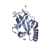

 PDBj
PDBj
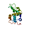
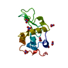
 Assembly


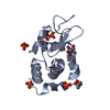
 Mass: 112.411 Da / Num. of mol.: 14 / Source method: obtained synthetically / Formula: Cd
 Mass: 18.015 Da / Num. of mol.: 107 / Source method: isolated from a natural source / Formula: H2O
 Sample preparation
 Processing