| Entry | Database: PDB / ID: 5azh
|
|---|
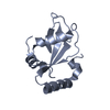
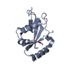
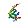
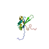
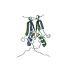
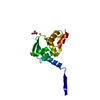
| Title | Crystal structure of LGG-2 fused with an EEEWEEL peptide |
|---|
 Components Components | EEEWEEL peptide,Protein lgg-2 |
|---|
 Keywords Keywords | PROTEIN BINDING / autophagy / ubiquitin-like |
|---|
| Function / homology |  Function and homology information Function and homology information
PINK1-PRKN Mediated Mitophagy / Receptor Mediated Mitophagy / Macroautophagy / KEAP1-NFE2L2 pathway / xenophagy / phosphatidylethanolamine binding / plasma membrane repair / cellular response to toxic substance / positive regulation of autophagosome maturation / cellular response to nitrogen starvation ...PINK1-PRKN Mediated Mitophagy / Receptor Mediated Mitophagy / Macroautophagy / KEAP1-NFE2L2 pathway / xenophagy / phosphatidylethanolamine binding / plasma membrane repair / cellular response to toxic substance / positive regulation of autophagosome maturation / cellular response to nitrogen starvation / autophagosome membrane / autophagosome maturation / autophagosome assembly / mitophagy / cytoplasmic vesicle / microtubule binding / defense response to Gram-positive bacterium / ubiquitin protein ligase binding / plasma membrane / cytoplasmSimilarity search - Function Autophagy protein Atg8 ubiquitin-like / Autophagy protein Atg8 ubiquitin like / Phosphatidylinositol 3-kinase Catalytic Subunit; Chain A, domain 1 / Ubiquitin-like (UB roll) / Ubiquitin-like domain superfamily / Roll / Alpha BetaSimilarity search - Domain/homology |
|---|
| Biological species | synthetic construct (others)
  Caenorhabditis elegans (invertebrata) Caenorhabditis elegans (invertebrata) |
|---|
| Method |  X-RAY DIFFRACTION / SYNCHROTRON / MOLECULAR REPLACEMENT / Resolution: 2.3 Å X-RAY DIFFRACTION / SYNCHROTRON / MOLECULAR REPLACEMENT / Resolution: 2.3 Å |
|---|
 Authors Authors | Watanabe, Y. / Fujioka, Y. / Noda, N.N. |
|---|
| Funding support |  Japan, 3items Japan, 3items | Organization | Grant number | Country |
|---|
| Ministry of Education, Culture, Sports, Science and Technology of Japan | 25111004 | Japan | | Ministry of Education, Culture, Sports, Science and Technology of Japan | 26870828 | Japan | | Japan Science and Technology Agency | CREST | Japan |
|
|---|
 Citation Citation | Journal: Mol.Cell / Year: 2015
Title: Structural Basis of the Differential Function of the Two C. elegans Atg8 Homologs, LGG-1 and LGG-2, in Autophagy.
Authors: Wu, F. / Watanabe, Y. / Guo, X.Y. / Qi, X. / Wang, P. / Zhao, H.Y. / Wang, Z. / Fujioka, Y. / Zhang, H. / Ren, J.Q. / Fang, T.C. / Shen, Y.X. / Feng, W. / Hu, J.J. / Noda, N.N. / Zhang, H. |
|---|
| History | | Deposition | Oct 5, 2015 | Deposition site: PDBJ / Processing site: PDBJ |
|---|
| Revision 1.0 | Dec 30, 2015 | Provider: repository / Type: Initial release |
|---|
| Revision 1.1 | Jan 1, 2020 | Group: Data collection / Database references / Derived calculations
Category: citation / diffrn_source / pdbx_struct_oper_list
Item: _citation.pdbx_database_id_PubMed / _citation.title ..._citation.pdbx_database_id_PubMed / _citation.title / _diffrn_source.pdbx_synchrotron_site / _pdbx_struct_oper_list.symmetry_operation |
|---|
| Revision 1.2 | Mar 20, 2024 | Group: Data collection / Database references / Derived calculations
Category: chem_comp_atom / chem_comp_bond ...chem_comp_atom / chem_comp_bond / database_2 / pdbx_struct_conn_angle / struct_conn
Item: _database_2.pdbx_DOI / _database_2.pdbx_database_accession ..._database_2.pdbx_DOI / _database_2.pdbx_database_accession / _pdbx_struct_conn_angle.ptnr1_auth_seq_id / _pdbx_struct_conn_angle.ptnr1_symmetry / _pdbx_struct_conn_angle.ptnr3_auth_seq_id / _pdbx_struct_conn_angle.ptnr3_symmetry / _pdbx_struct_conn_angle.value / _struct_conn.pdbx_dist_value / _struct_conn.ptnr2_auth_seq_id / _struct_conn.ptnr2_symmetry |
|---|
|
|---|
 Movie
Movie Controller


 Open data
 Basic information
 Structure visualization
 Downloads & links
 Other downloads
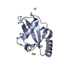


 PDBj
PDBj
 Assembly
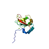


 Mass: 24.305 Da / Num. of mol.: 1 / Source method: obtained synthetically / Formula: Mg
 Mass: 18.015 Da / Num. of mol.: 35 / Source method: isolated from a natural source / Formula: H2O
 Sample preparation
 Processing