- PDB-5j8e: Crystal structure of human Hook3's conserved Hook domain -
+
Open data
ID or keywords:
Loading...
-
Basic information
Entry
Database: PDB / ID: 5j8e
Title
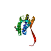
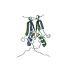
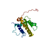
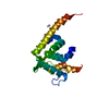
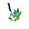
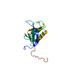
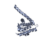
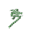
Crystal structure of human Hook3's conserved Hook domain
Components
Protein Hook homolog 3
Keywords
PROTEIN TRANSPORT / calponin homology Hook cargo adaptor
Function / homology
Function and homology information
FHF complex / interkinetic nuclear migration / protein localization to perinuclear region of cytoplasm / Golgi localization / microtubule anchoring at centrosome / dynein light chain binding / cytoskeleton-dependent intracellular transport / neuronal stem cell population maintenance / early endosome to late endosome transport / endosome organization ...FHF complex / interkinetic nuclear migration / protein localization to perinuclear region of cytoplasm / Golgi localization / microtubule anchoring at centrosome / dynein light chain binding / cytoskeleton-dependent intracellular transport / neuronal stem cell population maintenance / early endosome to late endosome transport / endosome organization / cis-Golgi network / protein localization to centrosome / dynein light intermediate chain binding / lysosome organization / endosome to lysosome transport / dynein intermediate chain binding / pericentriolar material / dynactin binding / cytoplasmic microtubule organization / negative regulation of neurogenesis / centriolar satellite / protein transport / microtubule binding / microtubule / centrosome / identical protein binding / cytoplasm Similarity search - Function
In the structure databanks used in Yorodumi, some data are registered as the other names, "COVID-19 virus" and "2019-nCoV". Here are the details of the virus and the list of structure data.
Jan 31, 2019. EMDB accession codes are about to change! (news from PDBe EMDB page)
EMDB accession codes are about to change! (news from PDBe EMDB page)
The allocation of 4 digits for EMDB accession codes will soon come to an end. Whilst these codes will remain in use, new EMDB accession codes will include an additional digit and will expand incrementally as the available range of codes is exhausted. The current 4-digit format prefixed with “EMD-” (i.e. EMD-XXXX) will advance to a 5-digit format (i.e. EMD-XXXXX), and so on. It is currently estimated that the 4-digit codes will be depleted around Spring 2019, at which point the 5-digit format will come into force.
The EM Navigator/Yorodumi systems omit the EMD- prefix.
Related info.:Q: What is EMD? / ID/Accession-code notation in Yorodumi/EM Navigator
Yorodumi is a browser for structure data from EMDB, PDB, SASBDB, etc.
This page is also the successor to EM Navigator detail page, and also detail information page/front-end page for Omokage search.
The word "yorodu" (or yorozu) is an old Japanese word meaning "ten thousand". "mi" (miru) is to see.
Related info.:EMDB / PDB / SASBDB / Comparison of 3 databanks / Yorodumi Search / Aug 31, 2016. New EM Navigator & Yorodumi / Yorodumi Papers / Jmol/JSmol / Function and homology information / Changes in new EM Navigator and Yorodumi
 Movie
Movie Controller


 Open data
 Basic information
 Components
 Keywords
 Function and homology information
 Homo sapiens (human)
 X-RAY DIFFRACTION /
  Authors
 United States, 1items
  Citation
 Structure visualization
 Downloads & links
 Other downloads


 PDBj
PDBj Assembly

 Mass: 18.015 Da / Num. of mol.: 181 / Source method: isolated from a natural source / Formula: H2O
 Sample preparation
 Processing