[English] 日本語
Yorodumi
- PDB-3udb: Crystal structure of SnRK2.6 -

+
Open data


ID or keywords:

Loading...

-
Basic information

Entry
Database: PDB / ID: 3udb
TitleCrystal structure of SnRK2.6
ComponentsSerine/threonine-protein kinase SRK2E
KeywordsTRANSFERASE / SnRK2.6 / kinase / ABA signaling pathway / ABI1
Function / homology
Function and homology information


cellular response to absence of light / regulation of anion channel activity / regulation of stomatal opening / unsaturated fatty acid biosynthetic process / cellular response to carbon dioxide / triglyceride biosynthetic process / positive regulation of abscisic acid-activated signaling pathway / sucrose metabolic process / regulation of stomatal closure / stomatal movement ...cellular response to absence of light / regulation of anion channel activity / regulation of stomatal opening / unsaturated fatty acid biosynthetic process / cellular response to carbon dioxide / triglyceride biosynthetic process / positive regulation of abscisic acid-activated signaling pathway / sucrose metabolic process / regulation of stomatal closure / stomatal movement / leaf development / regulation of stomatal movement / calcium-dependent protein serine/threonine kinase activity / response to water deprivation / response to abscisic acid / abscisic acid-activated signaling pathway / regulation of reactive oxygen species metabolic process / response to osmotic stress / response to salt stress / kinase activity / protein phosphatase binding / protein autophosphorylation / non-specific serine/threonine protein kinase / protein kinase activity / defense response to bacterium / protein serine kinase activity / ATP binding / identical protein binding / nucleus / plasma membrane / cytoplasm
Similarity search - Function
Transferase(Phosphotransferase) domain 1 / Transferase(Phosphotransferase); domain 1 / Phosphorylase Kinase; domain 1 / Phosphorylase Kinase; domain 1 / Serine/threonine-protein kinase, active site / Serine/Threonine protein kinases active-site signature. / Protein kinase domain / Serine/Threonine protein kinases, catalytic domain / Protein kinase, ATP binding site / Protein kinases ATP-binding region signature. ...Transferase(Phosphotransferase) domain 1 / Transferase(Phosphotransferase); domain 1 / Phosphorylase Kinase; domain 1 / Phosphorylase Kinase; domain 1 / Serine/threonine-protein kinase, active site / Serine/Threonine protein kinases active-site signature. / Protein kinase domain / Serine/Threonine protein kinases, catalytic domain / Protein kinase, ATP binding site / Protein kinases ATP-binding region signature. / Protein kinase domain profile. / Protein kinase domain / Protein kinase-like domain superfamily / 2-Layer Sandwich / Orthogonal Bundle / Mainly Alpha / Alpha Beta
Similarity search - Domain/homology
Serine/threonine-protein kinase SRK2E
Similarity search - Component
Biological speciesArabidopsis thaliana (thale cress)
MethodX-RAY DIFFRACTION / SYNCHROTRON / MOLECULAR REPLACEMENT / Resolution: 2.567 Å
AuthorsXie, T. / Ren, R. / Pang, Y. / Yan, C.
Citation
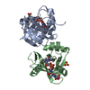
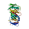
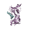
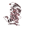
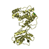
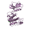
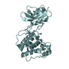
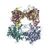
Journal: to be published
Title: Molecular mechanism for the inhibition of a critical component in the Arabidopsis thaliana abscisic acid signal transduction pathways, SnRK2.6, by the protein phosphatase ABI1
Authors: Xie, T. / Ren, R. / Pang, Y. / Yan, C.
History
DepositionOct 27, 2011Deposition site: RCSB / Processing site: PDBJ
Revision 1.0Nov 16, 2011Provider: repository / Type: Initial release
Revision 1.1Nov 1, 2023Group: Data collection / Database references / Refinement description
Category: chem_comp_atom / chem_comp_bond ...chem_comp_atom / chem_comp_bond / database_2 / pdbx_initial_refinement_model / struct_ref_seq_dif
Item: _database_2.pdbx_DOI / _database_2.pdbx_database_accession / _struct_ref_seq_dif.details

-
Structure visualization

Structure viewerMolecule:
MolmilJmol/JSmol

Downloads & links

-
Assembly

Deposited unit
A: Serine/threonine-protein kinase SRK2E
B: Serine/threonine-protein kinase SRK2E
C: Serine/threonine-protein kinase SRK2E
D: Serine/threonine-protein kinase SRK2E
E: Serine/threonine-protein kinase SRK2E
F: Serine/threonine-protein kinase SRK2E
hetero molecules


Theoretical massNumber of molelcules
Total (without water)216,09412
Polymers215,8816
Non-polymers2136
Water2,324129
1
A: Serine/threonine-protein kinase SRK2E
hetero molecules


Theoretical massNumber of molelcules
Total (without water)36,0162
Polymers35,9801
Non-polymers351
Water181
TypeNameSymmetry operationNumber
identity operation1_555x,y,z1
2
B: Serine/threonine-protein kinase SRK2E
hetero molecules


Theoretical massNumber of molelcules
Total (without water)36,0162
Polymers35,9801
Non-polymers351
Water181
TypeNameSymmetry operationNumber
identity operation1_555x,y,z1
3
C: Serine/threonine-protein kinase SRK2E
hetero molecules


Theoretical massNumber of molelcules
Total (without water)36,0162
Polymers35,9801
Non-polymers351
Water181
TypeNameSymmetry operationNumber
identity operation1_555x,y,z1
4
D: Serine/threonine-protein kinase SRK2E
hetero molecules


Theoretical massNumber of molelcules
Total (without water)36,0162
Polymers35,9801
Non-polymers351
Water181
TypeNameSymmetry operationNumber
identity operation1_555x,y,z1
5
E: Serine/threonine-protein kinase SRK2E
hetero molecules


Theoretical massNumber of molelcules
Total (without water)36,0162
Polymers35,9801
Non-polymers351
Water181
TypeNameSymmetry operationNumber
identity operation1_555x,y,z1
6
F: Serine/threonine-protein kinase SRK2E
hetero molecules


Theoretical massNumber of molelcules
Total (without water)36,0162
Polymers35,9801
Non-polymers351
Water181
TypeNameSymmetry operationNumber
identity operation1_555x,y,z1
7
A: Serine/threonine-protein kinase SRK2E
C: Serine/threonine-protein kinase SRK2E
D: Serine/threonine-protein kinase SRK2E
F: Serine/threonine-protein kinase SRK2E
hetero molecules


Theoretical massNumber of molelcules
Total (without water)144,0628
Polymers143,9214
Non-polymers1424
Water724
TypeNameSymmetry operationNumber
identity operation1_555x,y,z1
Buried area6530 Å2
ΔGint-59 kcal/mol
Surface area49540 Å2
MethodPISA
8
B: Serine/threonine-protein kinase SRK2E
E: Serine/threonine-protein kinase SRK2E
hetero molecules

B: Serine/threonine-protein kinase SRK2E
E: Serine/threonine-protein kinase SRK2E
hetero molecules


Theoretical massNumber of molelcules
Total (without water)144,0628
Polymers143,9214
Non-polymers1424
Water724
TypeNameSymmetry operationNumber
identity operation1_555x,y,z1
crystal symmetry operation2_556-x,y,-z+11
Buried area7090 Å2
ΔGint-71 kcal/mol
Surface area49820 Å2
MethodPISA
Unit cell
Length a, b, c (Å)228.094, 98.793, 113.103
Angle α, β, γ (deg.)90.00, 98.48, 90.00
Int Tables number5
Space group name H-MC121

-
Components

#1: Protein
Serine/threonine-protein kinase SRK2E / Protein OPEN STOMATA 1 / SNF1-related kinase 2.6 / SnRK2.6 / Serine/threonine-protein kinase OST1


Mass: 35980.152 Da / Num. of mol.: 6 / Fragment: residues 1-317 / Mutation: S7A/S29A/S43A/C131A/C137A/C159A/S166A/T176A
Source method: isolated from a genetically manipulated source
Source: (gene. exp.) Arabidopsis thaliana (thale cress) / Gene: SNRK2.6 / Production host: Escherichia coli (E. coli)
References: UniProt: Q940H6, non-specific serine/threonine protein kinase
#2: Chemical
ChemComp-CL / CHLORIDE ION / Chloride


Mass: 35.453 Da / Num. of mol.: 6 / Source method: obtained synthetically / Formula: Cl
#3: Water ChemComp-HOH / water / Water


Mass: 18.015 Da / Num. of mol.: 129 / Source method: isolated from a natural source / Formula: H2O

-
Experimental details

-
Experiment

ExperimentMethod: X-RAY DIFFRACTION / Number of used crystals: 1

-
Sample preparation

CrystalDensity Matthews: 2.92 Å3/Da / Density % sol: 57.87 %
Crystal growTemperature: 291 K / Method: vapor diffusion, hanging drop / pH: 8
Details: 0.1M Tris, 19% PEG4000, pH 8.0, VAPOR DIFFUSION, HANGING DROP, temperature 291K

-
Data collection

DiffractionMean temperature: 100 K
Diffraction sourceSource: SYNCHROTRON / Site: SSRF / Beamline: BL17U / Wavelength: 1.03887 Å
DetectorType: ADSC QUANTUM 315r / Detector: CCD / Date: Dec 14, 2010
RadiationMonochromator: Si 111 CHANNEL / Protocol: SINGLE WAVELENGTH / Monochromatic (M) / Laue (L): M / Scattering type: x-ray
Radiation wavelengthWavelength: 1.03887 Å / Relative weight: 1
ReflectionResolution: 2.567→40 Å / Num. all: 78743 / Num. obs: 78271 / % possible obs: 99.4 % / Observed criterion σ(F): 2 / Observed criterion σ(I): 2 / Biso Wilson estimate: 49.75 Å2
Reflection shellResolution: 2.58→2.67 Å / % possible all: 99.7

-
Processing

Software
NameVersionClassification
ADSCQuantumdata collection
PHASERphasing
PHENIX(phenix.refine: 1.7.1_743)refinement
HKL-2000data reduction
HKL-2000data scaling
RefinementMethod to determine structure: MOLECULAR REPLACEMENT
Starting model: 2FH9
Resolution: 2.567→37.076 Å / Occupancy max: 1 / Occupancy min: 0.45 / FOM work R set: 0.7649 / SU ML: 0.82 / σ(F): 1.34 / Phase error: 30.16 / Stereochemistry target values: ML
RfactorNum. reflection% reflectionSelection details
Rfree0.2594 3929 5.02 %RANDOM
Rwork0.2364 ---
all0.2378 78743 --
obs0.2376 78250 98.36 %-
Solvent computationShrinkage radii: 0.72 Å / VDW probe radii: 1 Å / Solvent model: FLAT BULK SOLVENT MODEL / Bsol: 36.958 Å2 / ksol: 0.291 e/Å3
Displacement parametersBiso max: 200.75 Å2 / Biso mean: 61.3777 Å2 / Biso min: 16.21 Å2
Baniso -1Baniso -2Baniso -3
1-3.2037 Å20 Å2-4.9171 Å2
2---9.396 Å2-0 Å2
3---6.7274 Å2
Refinement stepCycle: LAST / Resolution: 2.567→37.076 Å
ProteinNucleic acidLigandSolventTotal
Num. atoms13030 0 6 129 13165
Refine LS restraints
Refine-IDTypeDev idealNumber
X-RAY DIFFRACTIONf_bond_d0.0113338
X-RAY DIFFRACTIONf_angle_d1.59618033
X-RAY DIFFRACTIONf_chiral_restr0.1041992
X-RAY DIFFRACTIONf_plane_restr0.0142334
X-RAY DIFFRACTIONf_dihedral_angle_d20.4915035
LS refinement shell

Refine-ID: X-RAY DIFFRACTION / Total num. of bins used: 28

Resolution (Å)Rfactor RfreeNum. reflection RfreeRfactor RworkNum. reflection RworkNum. reflection all% reflection obs (%)
2.5667-2.5980.4232880.33171934202271
2.598-2.63090.39851290.350526642793100
2.6309-2.66550.38771430.350526872830100
2.6655-2.7020.39751310.33526692800100
2.702-2.74060.36891380.307326842822100
2.7406-2.78150.32761590.292726712830100
2.7815-2.82490.33821560.282726932849100
2.8249-2.87120.30151240.27726602784100
2.8712-2.92070.31081360.276626822818100
2.9207-2.97380.34861440.275726772821100
2.9738-3.03090.31071360.285926972833100
3.0309-3.09280.30371080.271726932801100
3.0928-3.160.31671350.27826892824100
3.16-3.23350.29761510.276126822833100
3.2335-3.31430.27741280.283526952823100
3.3143-3.40380.3181360.272527072843100
3.4038-3.50390.30441550.268626572812100
3.5039-3.61690.28821400.257426672807100
3.6169-3.74610.25991330.23252707284099
3.7461-3.89590.24241320.21542694282699
3.8959-4.0730.23781490.20392657280699
4.073-4.28750.20061390.18762692283199
4.2875-4.55560.21681520.17652683283599
4.5556-4.90660.1891670.17132624279199
4.9066-5.39910.25191600.21312702286299
5.3991-6.17720.25091470.24722684283199
6.1772-7.77080.20971580.20932680283899
7.7708-37.07940.21061550.21222690284597
Refinement TLS params.Method: refined / Origin x: -40.569 Å / Origin y: -8.5337 Å / Origin z: 32.6509 Å
111213212223313233
T0.221 Å20.0109 Å2-0.0185 Å2-0.1687 Å20.0063 Å2--0.2122 Å2
L0.1472 °20.0136 °20.0533 °2-0.1675 °20.067 °2--0.159 °2
S0.0118 Å °0.0171 Å °0.0303 Å °-0.0629 Å °0.0199 Å °-0.0351 Å °0.0268 Å °0.0019 Å °-0.0285 Å °
Refinement TLS group
IDRefine-IDRefine TLS-IDSelection detailsAuth asym-IDAuth seq-ID
1X-RAY DIFFRACTION1allA12 - 317
2X-RAY DIFFRACTION1allB12 - 317
3X-RAY DIFFRACTION1allC12 - 317
4X-RAY DIFFRACTION1allD12 - 317
5X-RAY DIFFRACTION1allE13 - 316
6X-RAY DIFFRACTION1allF12 - 317
7X-RAY DIFFRACTION1allD - B1 - 350
8X-RAY DIFFRACTION1allC - E1 - 318

+
About Yorodumi

-
News

-
Feb 9, 2022. New format data for meta-information of EMDB entries

New format data for meta-information of EMDB entries

  • Version 3 of the EMDB header file is now the official format.
  • The previous official version 1.9 will be removed from the archive.

Related info.:EMDB header

External links:wwPDB to switch to version 3 of the EMDB data model

-
Aug 12, 2020. Covid-19 info

Covid-19 info

URL: https://pdbj.org/emnavi/covid19.php

New page: Covid-19 featured information page in EM Navigator.

Related info.:Covid-19 info / Mar 5, 2020. Novel coronavirus structure data

+
Mar 5, 2020. Novel coronavirus structure data

Novel coronavirus structure data

Related info.:Yorodumi Speices / Aug 12, 2020. Covid-19 info

External links:COVID-19 featured content - PDBj / Molecule of the Month (242):Coronavirus Proteases

+
Jan 31, 2019. EMDB accession codes are about to change! (news from PDBe EMDB page)

EMDB accession codes are about to change! (news from PDBe EMDB page)

  • The allocation of 4 digits for EMDB accession codes will soon come to an end. Whilst these codes will remain in use, new EMDB accession codes will include an additional digit and will expand incrementally as the available range of codes is exhausted. The current 4-digit format prefixed with “EMD-” (i.e. EMD-XXXX) will advance to a 5-digit format (i.e. EMD-XXXXX), and so on. It is currently estimated that the 4-digit codes will be depleted around Spring 2019, at which point the 5-digit format will come into force.
  • The EM Navigator/Yorodumi systems omit the EMD- prefix.

Related info.:Q: What is EMD? / ID/Accession-code notation in Yorodumi/EM Navigator

External links:EMDB Accession Codes are Changing Soon! / Contact to PDBj

+
Jul 12, 2017. Major update of PDB

Major update of PDB

  • wwPDB released updated PDB data conforming to the new PDBx/mmCIF dictionary.
  • This is a major update changing the version number from 4 to 5, and with Remediation, in which all the entries are updated.
  • In this update, many items about electron microscopy experimental information are reorganized (e.g. em_software).
  • Now, EM Navigator and Yorodumi are based on the updated data.

External links:wwPDB Remediation / Enriched Model Files Conforming to OneDep Data Standards Now Available in the PDB FTP Archive

-
Yorodumi

Thousand views of thousand structures

  • Yorodumi is a browser for structure data from EMDB, PDB, SASBDB, etc.
  • This page is also the successor to EM Navigator detail page, and also detail information page/front-end page for Omokage search.
  • The word "yorodu" (or yorozu) is an old Japanese word meaning "ten thousand". "mi" (miru) is to see.

Related info.:EMDB / PDB / SASBDB / Comparison of 3 databanks / Yorodumi Search / Aug 31, 2016. New EM Navigator & Yorodumi / Yorodumi Papers / Jmol/JSmol / Function and homology information / Changes in new EM Navigator and Yorodumi

Read more