 Movie
Movie Controller

[English] 日本語
 Yorodumi
- PDB-3i2m: The Crystal Structure of PF-8, the DNA Polymerase Accessory Subun... -
+ Open data

- Basic information
| Entry | Database: PDB / ID: 3i2m | ||||||
|---|---|---|---|---|---|---|---|
| Title | The Crystal Structure of PF-8, the DNA Polymerase Accessory Subunit from Kaposi s Sarcoma-Associated Herpesvirus | ||||||
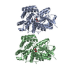
 Components Components | ORF59 | ||||||
 Keywords Keywords | REPLICATION / processivity | ||||||
| Function / homology | DNA polymerase processivity factor, herpesviridae / Herpes DNA replication accessory factor / Box / Proliferating Cell Nuclear Antigen / Proliferating Cell Nuclear Antigen - #10 / Alpha Beta / Core gene UL42 family protein Function and homology information Function and homology information | ||||||
| Biological species |   Human herpesvirus 8 Human herpesvirus 8 | ||||||
| Method |  X-RAY DIFFRACTION / SYNCHROTRON / SAD / Resolution: 2.81 Å X-RAY DIFFRACTION / SYNCHROTRON / SAD / Resolution: 2.81 Å | ||||||
 Authors Authors | Baltz, J.L. / Filman, D.J. / Ciustea, M. / Silverman, J.E.Y. / Lautenschlager, C.L. / Coen, D.M. / Ricciardi, R.P. / Hogle, J.M. | ||||||
 Citation Citation | Journal: J.Virol. / Year: 2009 Title: The crystal structure of PF-8, the DNA polymerase accessory subunit from Kaposi's sarcoma-associated herpesvirus. Authors: Baltz, J.L. / Filman, D.J. / Ciustea, M. / Silverman, J.E. / Lautenschlager, C.L. / Coen, D.M. / Ricciardi, R.P. / Hogle, J.M. | ||||||
| History |
|
- Structure visualization
| Structure viewer | Molecule: MolmilJmol/JSmol |
|---|
- Downloads & links
-Download
| PDBx/mmCIF format | 3i2m.cif.gz | 67.6 KB | Display | PDBx/mmCIF format |
|---|---|---|---|---|
| PDB format | pdb3i2m.ent.gz | 49.5 KB | Display | PDB format |
| PDBx/mmJSON format | 3i2m.json.gz | Tree view | PDBx/mmJSON format | |
| Others |  Other downloads Other downloads |
-Validation report
| Arichive directory | https://data.pdbj.org/pub/pdb/validation_reports/i2/3i2mftp://data.pdbj.org/pub/pdb/validation_reports/i2/3i2m | HTTPS FTP |
|---|
-Related structure data


-Links
 PDBj
PDBj- Assembly
| Deposited unit | 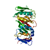
| ||||||||
|---|---|---|---|---|---|---|---|---|---|
| 1 | 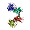
| ||||||||
| 2 |
| ||||||||
| Unit cell |
|
-Components
| #1: Protein | Mass: 33414.641 Da / Num. of mol.: 1 / Fragment: UNP residues 2-304 Source method: isolated from a genetically manipulated source Source: (gene. exp.) Human herpesvirus 8 / Production host:  |
|---|---|
| Has protein modification | Y |
-Experimental details
-Experiment
| Experiment | Method: X-RAY DIFFRACTION |
|---|
- Sample preparation
| Crystal | Density Matthews: 2.84 Å3/Da / Density % sol: 56.7 % |
|---|---|
| Crystal grow | Temperature: 295 K / Method: vapor diffusion, hanging drop / pH: 8.2 Details: 18% PEG 3350, 100 mM Tris HCl pH 8.2, 0.2 M lithium chloride, and 20 mM DTT, VAPOR DIFFUSION, HANGING DROP, temperature 295K |
-Data collection
| Diffraction source | Source: SYNCHROTRON / Site: APS  / Beamline: 24-ID-E / Wavelength: 0.97918 Å / Beamline: 24-ID-E / Wavelength: 0.97918 Å |
|---|---|
| Detector | Type: ADSC QUANTUM 315 / Detector: CCD / Date: Jan 1, 2008 |
| Radiation | Protocol: SINGLE WAVELENGTH / Monochromatic (M) / Laue (L): M / Scattering type: x-ray |
| Radiation wavelength | Wavelength: 0.97918 Å / Relative weight: 1 |
| Reflection | Resolution: 2.8→30 Å / Num. all: 10612 / Num. obs: 10103 / % possible obs: 95.2 % / Observed criterion σ(F): 0 / Observed criterion σ(I): -3 / Redundancy: 11.1 % / Rmerge(I) obs: 0.095 / Rsym value: 0.095 / Net I/σ(I): 18.012 |
| Reflection shell | Resolution: 2.8→2.9 Å / Redundancy: 0.049 % / Rmerge(I) obs: 0.492 / Mean I/σ(I) obs: 2.7 / Rsym value: 0.492 / % possible all: 62.1 |
- Processing
| Software |
| ||||||||||||||||||||||||||||||||||||||||||||||||||||||||||||||||||||||||||||||||||||||||||||||||||||||||||||||||||||||||||||||||||||||||||||||||||||||||||||||||||||||||||
|---|---|---|---|---|---|---|---|---|---|---|---|---|---|---|---|---|---|---|---|---|---|---|---|---|---|---|---|---|---|---|---|---|---|---|---|---|---|---|---|---|---|---|---|---|---|---|---|---|---|---|---|---|---|---|---|---|---|---|---|---|---|---|---|---|---|---|---|---|---|---|---|---|---|---|---|---|---|---|---|---|---|---|---|---|---|---|---|---|---|---|---|---|---|---|---|---|---|---|---|---|---|---|---|---|---|---|---|---|---|---|---|---|---|---|---|---|---|---|---|---|---|---|---|---|---|---|---|---|---|---|---|---|---|---|---|---|---|---|---|---|---|---|---|---|---|---|---|---|---|---|---|---|---|---|---|---|---|---|---|---|---|---|---|---|---|---|---|---|---|---|---|
| Refinement | Method to determine structure: SAD / Resolution: 2.81→29.15 Å / Cor.coef. Fo:Fc: 0.908 / Cor.coef. Fo:Fc free: 0.917 / SU B: 36.824 / SU ML: 0.324 / TLS residual ADP flag: LIKELY RESIDUAL / Cross valid method: THROUGHOUT / σ(F): 0 / ESU R: 1.284 / ESU R Free: 0.391 Stereochemistry target values: MAXIMUM LIKELIHOOD WITH PHASES
| ||||||||||||||||||||||||||||||||||||||||||||||||||||||||||||||||||||||||||||||||||||||||||||||||||||||||||||||||||||||||||||||||||||||||||||||||||||||||||||||||||||||||||
| Solvent computation | Ion probe radii: 0.8 Å / Shrinkage radii: 0.8 Å / VDW probe radii: 1.4 Å / Solvent model: BABINET MODEL WITH MASK | ||||||||||||||||||||||||||||||||||||||||||||||||||||||||||||||||||||||||||||||||||||||||||||||||||||||||||||||||||||||||||||||||||||||||||||||||||||||||||||||||||||||||||
| Displacement parameters | Biso mean: 31.36 Å2
| ||||||||||||||||||||||||||||||||||||||||||||||||||||||||||||||||||||||||||||||||||||||||||||||||||||||||||||||||||||||||||||||||||||||||||||||||||||||||||||||||||||||||||
| Refinement step | Cycle: LAST / Resolution: 2.81→29.15 Å
| ||||||||||||||||||||||||||||||||||||||||||||||||||||||||||||||||||||||||||||||||||||||||||||||||||||||||||||||||||||||||||||||||||||||||||||||||||||||||||||||||||||||||||
| Refine LS restraints |
| ||||||||||||||||||||||||||||||||||||||||||||||||||||||||||||||||||||||||||||||||||||||||||||||||||||||||||||||||||||||||||||||||||||||||||||||||||||||||||||||||||||||||||
| LS refinement shell | Resolution: 2.81→2.956 Å / Total num. of bins used: 10
| ||||||||||||||||||||||||||||||||||||||||||||||||||||||||||||||||||||||||||||||||||||||||||||||||||||||||||||||||||||||||||||||||||||||||||||||||||||||||||||||||||||||||||
| Refinement TLS params. | Method: refined / Origin x: 27.7329 Å / Origin y: -14.1581 Å / Origin z: 203.4016 Å
|
