 Movie
Movie Controller

[English] 日本語
 Yorodumi
- PDB-3r9x: Crystal structure of Era in complex with MgGDPNP, nucleotides 150... -
+ Open data

- Basic information
| Entry | Database: PDB / ID: 3r9x | ||||||
|---|---|---|---|---|---|---|---|
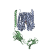
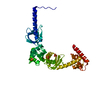
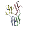
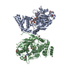
| Title | Crystal structure of Era in complex with MgGDPNP, nucleotides 1506-1542 of 16S ribosomal RNA, and KsgA | ||||||
 Components Components |
| ||||||
 Keywords Keywords | HYDROLASE/TRANSFERASE/RNA / GTPase / KH domain / ribosome / biogenesis / GTP / 16S ribosomal RNA / GTP hydrolysis / HYDROLASE-TRANSFERASE-RNA complex | ||||||
| Function / homology |  Function and homology information Function and homology information16S rRNA (adenine1518-N6/adenine1519-N6)-dimethyltransferase / 16S rRNA (adenine(1518)-N(6)/adenine(1519)-N(6))-dimethyltransferase activity / rRNA (adenine-N6,N6-)-dimethyltransferase activity / rRNA methylation / ribosomal small subunit binding / ribosomal small subunit assembly / small ribosomal subunit rRNA binding / rRNA binding / GTPase activity / GTP binding ...16S rRNA (adenine1518-N6/adenine1519-N6)-dimethyltransferase / 16S rRNA (adenine(1518)-N(6)/adenine(1519)-N(6))-dimethyltransferase activity / rRNA (adenine-N6,N6-)-dimethyltransferase activity / rRNA methylation / ribosomal small subunit binding / ribosomal small subunit assembly / small ribosomal subunit rRNA binding / rRNA binding / GTPase activity / GTP binding / RNA binding / plasma membrane / cytosol Similarity search - Function | ||||||
| Biological species |   Aquifex aeolicus (bacteria) Aquifex aeolicus (bacteria) | ||||||
| Method |  X-RAY DIFFRACTION / SYNCHROTRON / MOLECULAR REPLACEMENT / molecular replacement / Resolution: 2.8 Å X-RAY DIFFRACTION / SYNCHROTRON / MOLECULAR REPLACEMENT / molecular replacement / Resolution: 2.8 Å | ||||||
 Authors Authors | Tu, C. / Ji, X. | ||||||
 Citation Citation | Journal: Proc.Natl.Acad.Sci.USA / Year: 2011 Title: The Era GTPase recognizes the GAUCACCUCC sequence and binds helix 45 near the 3' end of 16S rRNA. Authors: Tu, C. / Zhou, X. / Tarasov, S.G. / Tropea, J.E. / Austin, B.P. / Waugh, D.S. / Court, D.L. / Ji, X. #1: Journal: Proc.Natl.Acad.Sci.USA / Year: 2009Title: Structure of ERA in complex with the 3' end of 16S rRNA: Implications for ribosome biogenesis Authors: Tu, C. / Zhou, X. / Tropea, J.E. / Austin, B.P. / Waugh, D.S. / Court, L.D. / Ji, X. #2: Journal: Proc.Natl.Acad.Sci.USA / Year: 1999Title: Crystal structure of ERA: A GTPase-dependent cell cycle regulator containing an RNA binding motif Authors: Chen, X. / Court, L.D. / Ji, X. | ||||||
| History |
|
- Structure visualization
| Structure viewer | Molecule: MolmilJmol/JSmol |
|---|
- Downloads & links
-Download
| PDBx/mmCIF format | 3r9x.cif.gz | 278.5 KB | Display | PDBx/mmCIF format |
|---|---|---|---|---|
| PDB format | pdb3r9x.ent.gz | 220.2 KB | Display | PDB format |
| PDBx/mmJSON format | 3r9x.json.gz | Tree view | PDBx/mmJSON format | |
| Others |  Other downloads Other downloads |
-Validation report
| Arichive directory | https://data.pdbj.org/pub/pdb/validation_reports/r9/3r9xftp://data.pdbj.org/pub/pdb/validation_reports/r9/3r9x | HTTPS FTP |
|---|
-Related structure data
| Related structure data |  3r9wC  3ftfS  3ievS S: Starting model for refinement C: citing same article ( |
|---|---|
| Similar structure data |


-Links
 PDBj
PDBj


- Assembly
| Deposited unit | 
| ||||||||
|---|---|---|---|---|---|---|---|---|---|
| 1 |
| ||||||||
| Unit cell |
|
-Components
-Protein , 2 types, 2 molecules AB
| #1: Protein | Mass: 34765.027 Da / Num. of mol.: 1 Source method: isolated from a genetically manipulated source Source: (gene. exp.) Aquifex aeolicus (bacteria) / Gene: aq_1994, era, era1 / Plasmid: pDONR201 / Production host: |
|---|---|
| #2: Protein | Mass: 28397.281 Da / Num. of mol.: 1 Source method: isolated from a genetically manipulated source Source: (gene. exp.) Aquifex aeolicus (bacteria) / Gene: aq_1816, ksgA, rsmA / Plasmid: pDONR201 / Production host: References: UniProt: O67680, 16S rRNA (adenine1518-N6/adenine1519-N6)-dimethyltransferase |
-RNA chain , 1 types, 1 molecules C
| #3: RNA chain | Mass: 11211.701 Da / Num. of mol.: 1 / Source method: obtained synthetically Details: This sequence occurs naturally in Aquifex aeolicus except for U1506 that is replaced with C1506. |
|---|
-Non-polymers , 5 types, 89 molecules 


| #4: Chemical | ChemComp-MG /  Mass: 24.305 Da / Num. of mol.: 1 / Source method: obtained synthetically / Formula: Mg Mass: 24.305 Da / Num. of mol.: 1 / Source method: obtained synthetically / Formula: Mg | ||||
|---|---|---|---|---|---|
| #5: Chemical | ChemComp-GNP /  Mass: 522.196 Da / Num. of mol.: 1 / Source method: obtained synthetically / Formula: C10H17N6O13P3 Mass: 522.196 Da / Num. of mol.: 1 / Source method: obtained synthetically / Formula: C10H17N6O13P3Comment: GppNHp, GMPPNP, energy-carrying molecule analogue*YM | ||||
| #6: Chemical |  Mass: 118.174 Da / Num. of mol.: 2 / Source method: obtained synthetically / Formula: C6H14O2 / Comment: precipitant*YM Mass: 118.174 Da / Num. of mol.: 2 / Source method: obtained synthetically / Formula: C6H14O2 / Comment: precipitant*YM#7: Chemical |  Mass: 59.044 Da / Num. of mol.: 2 / Source method: obtained synthetically / Formula: C2H3O2 Mass: 59.044 Da / Num. of mol.: 2 / Source method: obtained synthetically / Formula: C2H3O2#8: Water | ChemComp-HOH / | Mass: 18.015 Da / Num. of mol.: 83 / Source method: isolated from a natural source / Formula: H2O |
-Details
| Has protein modification | Y |
|---|
-Experimental details
-Experiment
| Experiment | Method: X-RAY DIFFRACTION / Number of used crystals: 1 |
|---|
- Sample preparation
| Crystal | Density Matthews: 3.24 Å3/Da / Density % sol: 62 % |
|---|---|
| Crystal grow | Temperature: 292 K / Method: vapor diffusion, sitting drop / pH: 5 Details: 0.1M sodium acetate, 20% (v/v) MPD, pH 5.0, VAPOR DIFFUSION, SITTING DROP, temperature 292K |
-Data collection
| Diffraction | Mean temperature: 100 K | |||||||||||||||||||||||||||||||||||||||||||||||||||||||||||||||||||||||||||||
|---|---|---|---|---|---|---|---|---|---|---|---|---|---|---|---|---|---|---|---|---|---|---|---|---|---|---|---|---|---|---|---|---|---|---|---|---|---|---|---|---|---|---|---|---|---|---|---|---|---|---|---|---|---|---|---|---|---|---|---|---|---|---|---|---|---|---|---|---|---|---|---|---|---|---|---|---|---|---|
| Diffraction source | Source: SYNCHROTRON / Site: APS  / Beamline: 22-BM / Wavelength: 1 Å / Beamline: 22-BM / Wavelength: 1 Å | |||||||||||||||||||||||||||||||||||||||||||||||||||||||||||||||||||||||||||||
| Detector | Type: MARMOSAIC 225 mm CCD / Detector: CCD / Date: Oct 29, 2008 / Details: mirrors | |||||||||||||||||||||||||||||||||||||||||||||||||||||||||||||||||||||||||||||
| Radiation | Monochromator: GRAPHITE / Protocol: SINGLE WAVELENGTH / Monochromatic (M) / Laue (L): M / Scattering type: x-ray | |||||||||||||||||||||||||||||||||||||||||||||||||||||||||||||||||||||||||||||
| Radiation wavelength | Wavelength: 1 Å / Relative weight: 1 | |||||||||||||||||||||||||||||||||||||||||||||||||||||||||||||||||||||||||||||
| Reflection | Resolution: 2.8→30 Å / Num. all: 23217 / Num. obs: 23217 / % possible obs: 98.4 % / Observed criterion σ(F): -6 / Observed criterion σ(I): -3 / Redundancy: 4.8 % / Biso Wilson estimate: 56.8 Å2 / Rmerge(I) obs: 0.097 / Χ2: 1.024 / Net I/σ(I): 9.4 | |||||||||||||||||||||||||||||||||||||||||||||||||||||||||||||||||||||||||||||
| Reflection shell | Diffraction-ID: 1
|
-Phasing
| Phasing | Method: molecular replacement |
|---|
- Processing
| Software |
| ||||||||||||||||||||||||||||||||||||||||||||||||||||||||||||||||
|---|---|---|---|---|---|---|---|---|---|---|---|---|---|---|---|---|---|---|---|---|---|---|---|---|---|---|---|---|---|---|---|---|---|---|---|---|---|---|---|---|---|---|---|---|---|---|---|---|---|---|---|---|---|---|---|---|---|---|---|---|---|---|---|---|---|
| Refinement | Method to determine structure: MOLECULAR REPLACEMENT Starting model: 3IEV, 3FTF Resolution: 2.8→28.686 Å / Occupancy max: 1 / Occupancy min: 0 / SU ML: 0.37 / Cross valid method: THROUGHOUT / σ(F): 0.09 / Stereochemistry target values: ML
| ||||||||||||||||||||||||||||||||||||||||||||||||||||||||||||||||
| Solvent computation | Shrinkage radii: 0.9 Å / VDW probe radii: 1.11 Å / Solvent model: FLAT BULK SOLVENT MODEL / Bsol: 44.228 Å2 / ksol: 0.284 e/Å3 | ||||||||||||||||||||||||||||||||||||||||||||||||||||||||||||||||
| Displacement parameters | Biso max: 250.4 Å2 / Biso mean: 75.835 Å2 / Biso min: 12.88 Å2
| ||||||||||||||||||||||||||||||||||||||||||||||||||||||||||||||||
| Refinement step | Cycle: LAST / Resolution: 2.8→28.686 Å
| ||||||||||||||||||||||||||||||||||||||||||||||||||||||||||||||||
| Refine LS restraints |
| ||||||||||||||||||||||||||||||||||||||||||||||||||||||||||||||||
| LS refinement shell | Refine-ID: X-RAY DIFFRACTION / Total num. of bins used: 7
|
