 Movie
Movie Controller

[English] 日本語
 Yorodumi
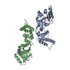
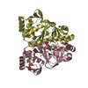
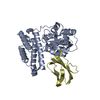
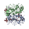
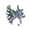
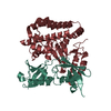
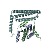
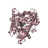
- PDB-3ftf: Crystal structure of A. aeolicus KsgA in complex with RNA and SAH -
+ Open data

- Basic information
| Entry | Database: PDB / ID: 3ftf | ||||||
|---|---|---|---|---|---|---|---|
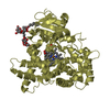
| Title | Crystal structure of A. aeolicus KsgA in complex with RNA and SAH | ||||||
 Components Components |
| ||||||
 Keywords Keywords | TRANSFERASE/RNA / KsgA / Rossmann-like fold / RNA Methyltransferase / MTase / RNA / Antibiotic resistance / Methyltransferase / RNA-binding / rRNA processing / S-adenosyl-L-methionine / Transferase / TRANSFERASE-RNA COMPLEX | ||||||
| Function / homology |  Function and homology information Function and homology information16S rRNA (adenine1518-N6/adenine1519-N6)-dimethyltransferase / 16S rRNA (adenine(1518)-N(6)/adenine(1519)-N(6))-dimethyltransferase activity / rRNA (adenine-N6,N6-)-dimethyltransferase activity / rRNA methylation / RNA binding / cytosol Similarity search - Function | ||||||
| Biological species |   Aquifex aeolicus (bacteria) Aquifex aeolicus (bacteria) | ||||||
| Method |  X-RAY DIFFRACTION / SYNCHROTRON / MOLECULAR REPLACEMENT / Resolution: 2.8 Å X-RAY DIFFRACTION / SYNCHROTRON / MOLECULAR REPLACEMENT / Resolution: 2.8 Å | ||||||
 Authors Authors | Tu, C. / Ji, X. | ||||||
 Citation Citation | Journal: Structure / Year: 2009 Title: Structural Basis for Binding of RNA and Cofactor by a KsgA Methyltransferase. Authors: Tu, C. / Tropea, J.E. / Austin, B.P. / Court, D.L. / Waugh, D.S. / Ji, X. | ||||||
| History |
|
- Structure visualization
| Structure viewer | Molecule: MolmilJmol/JSmol |
|---|
- Downloads & links
-Download
| PDBx/mmCIF format | 3ftf.cif.gz | 89.7 KB | Display | PDBx/mmCIF format |
|---|---|---|---|---|
| PDB format | pdb3ftf.ent.gz | 62.9 KB | Display | PDB format |
| PDBx/mmJSON format | 3ftf.json.gz | Tree view | PDBx/mmJSON format | |
| Others |  Other downloads Other downloads |
-Validation report
| Arichive directory | https://data.pdbj.org/pub/pdb/validation_reports/ft/3ftfftp://data.pdbj.org/pub/pdb/validation_reports/ft/3ftf | HTTPS FTP |
|---|
-Related structure data
| Related structure data | 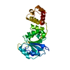 3ftcSC 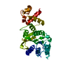 3ftdC  3fteC S: Starting model for refinement C: citing same article ( |
|---|---|
| Similar structure data |


-Links
 PDBj
PDBj


- Assembly
| Deposited unit | 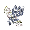
| ||||||||
|---|---|---|---|---|---|---|---|---|---|
| 1 |
| ||||||||
| Unit cell |
| ||||||||
| Details | The biological assembly corresponds to the content of the asymmetric unit. |
-Components
| #1: Protein | Mass: 28484.355 Da / Num. of mol.: 1 Source method: isolated from a genetically manipulated source Details: genomic DNA / Source: (gene. exp.) Aquifex aeolicus (bacteria) / Strain: VF5 / Gene: aq_1816, ksgA / Plasmid: pBA1939 / Production host: References: UniProt: O67680, Transferases; Transferring one-carbon groups; Methyltransferases | ||||||||
|---|---|---|---|---|---|---|---|---|---|
| #2: RNA chain | Mass: 7113.291 Da / Num. of mol.: 2 / Source method: obtained synthetically / Details: 16S rRNA #3: Chemical | ChemComp-SAH / |   Mass: 384.411 Da / Num. of mol.: 1 / Source method: obtained synthetically / Formula: C14H20N6O5S Mass: 384.411 Da / Num. of mol.: 1 / Source method: obtained synthetically / Formula: C14H20N6O5S#4: Chemical | ChemComp-K / |   Mass: 39.098 Da / Num. of mol.: 1 / Source method: obtained synthetically / Formula: K Mass: 39.098 Da / Num. of mol.: 1 / Source method: obtained synthetically / Formula: K#5: Water | ChemComp-HOH / |  Mass: 18.015 Da / Num. of mol.: 36 / Source method: isolated from a natural source / Formula: H2O Mass: 18.015 Da / Num. of mol.: 36 / Source method: isolated from a natural source / Formula: H2OHas protein modification | Y | |
-Experimental details
-Experiment
| Experiment | Method: X-RAY DIFFRACTION / Number of used crystals: 1 |
|---|
- Sample preparation
| Crystal | Density Matthews: 1.9 Å3/Da / Density % sol: 35.36 % | ||||||||||||||||||||
|---|---|---|---|---|---|---|---|---|---|---|---|---|---|---|---|---|---|---|---|---|---|
| Crystal grow | Temperature: 292 K / Method: vapor diffusion, sitting drop / pH: 7.2 Details: 0.2 M KF, 40% MPD, pH 7.2, VAPOR DIFFUSION, SITTING DROP, temperature 292K | ||||||||||||||||||||
| Components of the solutions |
|
-Data collection
| Diffraction | Mean temperature: 100 K |
|---|---|
| Diffraction source | Source: SYNCHROTRON / Site: APS  / Beamline: 22-BM / Wavelength: 1 Å / Beamline: 22-BM / Wavelength: 1 Å |
| Detector | Type: MARMOSAIC 225 mm CCD / Detector: CCD / Date: Oct 6, 2007 / Details: mirrors |
| Radiation | Monochromator: Si(111) Rosenbaum-Rock double crystal / Protocol: SINGLE WAVELENGTH / Monochromatic (M) / Laue (L): M / Scattering type: x-ray |
| Radiation wavelength | Wavelength: 1 Å / Relative weight: 1 |
| Reflection | Resolution: 2.78→30 Å / Num. all: 6848 / Num. obs: 6848 / % possible obs: 84.2 % / Observed criterion σ(I): -3 / Redundancy: 3.9 % / Biso Wilson estimate: 33.7 Å2 / Rmerge(I) obs: 0.144 / Net I/σ(I): 9 |
| Reflection shell | Resolution: 2.78→2.88 Å / Redundancy: 2.1 % / Rmerge(I) obs: 0.397 / Mean I/σ(I) obs: 2.4 / Num. unique all: 410 / Rsym value: 0.342 / % possible all: 49.9 |
- Processing
| Software |
| ||||||||||||||||||||||||||||||||||||
|---|---|---|---|---|---|---|---|---|---|---|---|---|---|---|---|---|---|---|---|---|---|---|---|---|---|---|---|---|---|---|---|---|---|---|---|---|---|
| Refinement | Method to determine structure: MOLECULAR REPLACEMENT Starting model: PDB entry 3FTC Resolution: 2.8→28.33 Å / Isotropic thermal model: Isotropic / Cross valid method: THROUGHOUT / Stereochemistry target values: Engh & Huber Details: 1. THE EFFECTIVE RESOLUTION FOR THIS STRUCTURE IS 3.05 ANGSTROM, AT WHICH THE COMPLETENESS OF X-RAY DATA > 93% AND THE OBSERVABLE DATA > 70% FOR HIGHEST RESOLUTION SHELL. 2. HYDROGENS HAVE ...Details: 1. THE EFFECTIVE RESOLUTION FOR THIS STRUCTURE IS 3.05 ANGSTROM, AT WHICH THE COMPLETENESS OF X-RAY DATA > 93% AND THE OBSERVABLE DATA > 70% FOR HIGHEST RESOLUTION SHELL. 2. HYDROGENS HAVE BEEN ADDED IN THE RIDING POSITIONS
| ||||||||||||||||||||||||||||||||||||
| Displacement parameters | Biso mean: 28.678 Å2
| ||||||||||||||||||||||||||||||||||||
| Refine analyze | Luzzati sigma a obs: 0.43 Å | ||||||||||||||||||||||||||||||||||||
| Refinement step | Cycle: LAST / Resolution: 2.8→28.33 Å
| ||||||||||||||||||||||||||||||||||||
| Refine LS restraints |
| ||||||||||||||||||||||||||||||||||||
| LS refinement shell | Refine-ID: X-RAY DIFFRACTION / Total num. of bins used: 5
|
