 Movie
Movie Controller


+ Open data

- Basic information
| Entry | Database: PDB / ID: 6xwx | ||||||
|---|---|---|---|---|---|---|---|
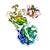
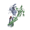
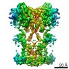
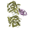
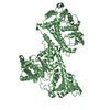
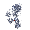
| Title | Crystal structure of the Tof1-Csm3 complex | ||||||
 Components Components |
| ||||||
 Keywords Keywords | REPLICATION / Fork Protection Complex / Replisome / Cohesion establishment / S phase checkpoint / Armadillo repeat | ||||||
| Function / homology |  Function and homology information Function and homology informationreplication fork protection complex / replication fork arrest / DNA replication checkpoint signaling / negative regulation of DNA replication / replication fork processing / meiotic cell cycle / DNA damage response / DNA binding / nucleus Similarity search - Function | ||||||
| Biological species |  Chaetomium thermophilum (fungus) Chaetomium thermophilum (fungus) | ||||||
| Method |  X-RAY DIFFRACTION / SYNCHROTRON / MOLECULAR REPLACEMENT / Resolution: 3.1 Å X-RAY DIFFRACTION / SYNCHROTRON / MOLECULAR REPLACEMENT / Resolution: 3.1 Å | ||||||
 Authors Authors | Grabarczyk, D.B. | ||||||
| Funding support |  Germany, 1items Germany, 1items
| ||||||
 Citation Citation | Journal: Nucleic Acids Res. / Year: 2020 Title: Crystal structure and interactions of the Tof1-Csm3 (Timeless-Tipin) fork protection complex. Authors: Grabarczyk, D.B. | ||||||
| History |
|
- Structure visualization
| Structure viewer | Molecule: MolmilJmol/JSmol |
|---|
- Downloads & links
-Download
| PDBx/mmCIF format | 6xwx.cif.gz | 387.6 KB | Display | PDBx/mmCIF format |
|---|---|---|---|---|
| PDB format | pdb6xwx.ent.gz | 318.7 KB | Display | PDB format |
| PDBx/mmJSON format | 6xwx.json.gz | Tree view | PDBx/mmJSON format | |
| Others |  Other downloads Other downloads |
-Validation report
| Arichive directory | https://data.pdbj.org/pub/pdb/validation_reports/xw/6xwxftp://data.pdbj.org/pub/pdb/validation_reports/xw/6xwx | HTTPS FTP |
|---|
-Related structure data
| Related structure data | 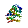 5mqiS S: Starting model for refinement |
|---|---|
| Similar structure data |


-Links
 PDBj
PDBj- Assembly
| Deposited unit | 
| ||||||||
|---|---|---|---|---|---|---|---|---|---|
| 1 | 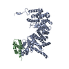
| ||||||||
| 2 | 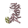
| ||||||||
| 3 | 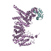
| ||||||||
| Unit cell |
|
-Components
| #1: Protein | Mass: 69540.414 Da / Num. of mol.: 3 Source method: isolated from a genetically manipulated source Source: (gene. exp.) Chaetomium thermophilum (strain DSM 1495 / CBS 144.50 / IMI 039719) (fungus)Gene: CTHT_0029130 / Production host:  #2: Protein | Mass: 12895.146 Da / Num. of mol.: 3 Source method: isolated from a genetically manipulated source Source: (gene. exp.) Chaetomium thermophilum (strain DSM 1495 / CBS 144.50 / IMI 039719) (fungus)Gene: CTHT_0011090 / Production host: |
|---|
-Experimental details
-Experiment
| Experiment | Method: X-RAY DIFFRACTION / Number of used crystals: 1 |
|---|
- Sample preparation
| Crystal | Density Matthews: 2.9 Å3/Da / Density % sol: 57.6 % |
|---|---|
| Crystal grow | Temperature: 293 K / Method: vapor diffusion, sitting drop Details: 0.2 M Potassium Acetate, 0.1 M Tris-HCl pH 7.5-8.5, 4-8% PEG 20000 |
-Data collection
| Diffraction | Mean temperature: 100 K / Serial crystal experiment: N |
|---|---|
| Diffraction source | Source: SYNCHROTRON / Site: PETRA III, EMBL c/o DESY / Beamline: P14 (MX2) / Wavelength: 0.98 Å |
| Detector | Type: DECTRIS EIGER X 16M / Detector: PIXEL / Date: May 27, 2019 |
| Radiation | Protocol: SINGLE WAVELENGTH / Monochromatic (M) / Laue (L): M / Scattering type: x-ray |
| Radiation wavelength | Wavelength: 0.98 Å / Relative weight: 1 |
| Reflection | Resolution: 3.09→76.74 Å / Num. obs: 27164 / % possible obs: 89.8 % / Redundancy: 7 % / CC1/2: 0.996 / Rpim(I) all: 0.078 / Net I/σ(I): 7.1 |
| Reflection shell | Resolution: 3.09→3.524 Å / Mean I/σ(I) obs: 1.2 / Num. unique obs: 1359 / CC1/2: 0.365 / Rpim(I) all: 0.737 |
- Processing
| Software |
| ||||||||||||||||||||
|---|---|---|---|---|---|---|---|---|---|---|---|---|---|---|---|---|---|---|---|---|---|
| Refinement | Method to determine structure: MOLECULAR REPLACEMENT Starting model: 5MQI Resolution: 3.1→45.73 Å / Cor.coef. Fo:Fc: 0.909 / Cor.coef. Fo:Fc free: 0.895 / Cross valid method: THROUGHOUT / SU Rfree Blow DPI: 0.741
| ||||||||||||||||||||
| Displacement parameters | Biso mean: 123.41 Å2
| ||||||||||||||||||||
| Refine analyze | Luzzati coordinate error obs: 0.64 Å | ||||||||||||||||||||
| Refinement step | Cycle: LAST / Resolution: 3.1→45.73 Å
| ||||||||||||||||||||
| LS refinement shell | Resolution: 3.1→3.39 Å
|
