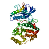
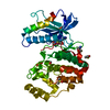
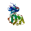
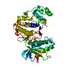
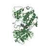
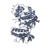
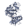
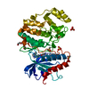
Mass: 43858.801 Da / Num. of mol.: 1 Source method: isolated from a genetically manipulated source Details: The recombinant version of the N-terminal domain of human Timeless used for X-ray crystal structure determination carried an internal deletion of amino acids 239 to 330, which were replaced ...Details: The recombinant version of the N-terminal domain of human Timeless used for X-ray crystal structure determination carried an internal deletion of amino acids 239 to 330, which were replaced with the artificial linker sequence GSTGST. Source: (gene. exp.) Homo sapiens (human) / Gene: TIMELESS, TIM, TIM1, TIMELESS1 / Production host: Escherichia coli (E. coli) / References: UniProt: Q9UNS1
Mass: 18.015 Da / Num. of mol.: 161 / Source method: isolated from a natural source / Formula: H2O
-
Experimental details
-
Experiment
Experiment
Method: X-RAY DIFFRACTION / Number of used crystals: 1
-
Sample preparation
Crystal
Density Matthews: 2.56 Å3/Da / Density % sol: 51.93 %
Crystal grow
Temperature: 292 K / Method: vapor diffusion, sitting drop Details: reservoir solution: 20% (w/v) PEG 3350, 200 mM Na2SO4 protein solution: 8.5 mg/mL, 150 mM KCl, 25 mM Heps pH 7.2, 1 mM TCEP 200 nL protein solution and 200 nL of reservoir solution were mixed.
In the structure databanks used in Yorodumi, some data are registered as the other names, "COVID-19 virus" and "2019-nCoV". Here are the details of the virus and the list of structure data.
Jan 31, 2019. EMDB accession codes are about to change! (news from PDBe EMDB page)
EMDB accession codes are about to change! (news from PDBe EMDB page)
The allocation of 4 digits for EMDB accession codes will soon come to an end. Whilst these codes will remain in use, new EMDB accession codes will include an additional digit and will expand incrementally as the available range of codes is exhausted. The current 4-digit format prefixed with “EMD-” (i.e. EMD-XXXX) will advance to a 5-digit format (i.e. EMD-XXXXX), and so on. It is currently estimated that the 4-digit codes will be depleted around Spring 2019, at which point the 5-digit format will come into force.
The EM Navigator/Yorodumi systems omit the EMD- prefix.
Related info.:Q: What is EMD? / ID/Accession-code notation in Yorodumi/EM Navigator
Yorodumi is a browser for structure data from EMDB, PDB, SASBDB, etc.
This page is also the successor to EM Navigator detail page, and also detail information page/front-end page for Omokage search.
The word "yorodu" (or yorozu) is an old Japanese word meaning "ten thousand". "mi" (miru) is to see.
Related info.:EMDB / PDB / SASBDB / Comparison of 3 databanks / Yorodumi Search / Aug 31, 2016. New EM Navigator & Yorodumi / Yorodumi Papers / Jmol/JSmol / Function and homology information / Changes in new EM Navigator and Yorodumi
 Movie
Movie Controller


 Open data
 Basic information
 Components
 Keywords
 Function and homology information
 Homo sapiens (human)
 X-RAY DIFFRACTION /
  Authors
 United Kingdom, 1items
  Citation
 Structure visualization
 Downloads & links
 Other downloads
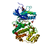

 PDBj
PDBj Assembly
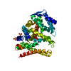


 Mass: 96.063 Da / Num. of mol.: 4 / Source method: obtained synthetically / Formula: SO4
 Mass: 18.015 Da / Num. of mol.: 161 / Source method: isolated from a natural source / Formula: H2O
 Sample preparation
 / Beamline: PROXIMA 1 / Wavelength: 0.97857 Å
 Processing