 Movie
Movie Controller

[English] 日本語
 Yorodumi
- PDB-5es9: Crystal structure of the LgrA initiation module in the formylatio... -
+ Open data

- Basic information
| Entry | Database: PDB / ID: 5es9 | ||||||
|---|---|---|---|---|---|---|---|
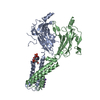
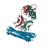
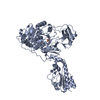
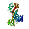
| Title | Crystal structure of the LgrA initiation module in the formylation state | ||||||
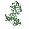
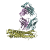
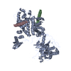
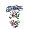
 Components Components | Linear gramicidin synthetase subunit A | ||||||
 Keywords Keywords | LIGASE / NRPS / formylation domain / adenylation domain / peptidyl carrier protein / initiation module | ||||||
| Function / homology |  Function and homology information Function and homology informationamino acid activation for nonribosomal peptide biosynthetic process / secondary metabolite biosynthetic process / lipid biosynthetic process / ligase activity / phosphopantetheine binding / antibiotic biosynthetic process / cytoplasm Similarity search - Function | ||||||
| Biological species |  Brevibacillus parabrevis (bacteria) Brevibacillus parabrevis (bacteria) | ||||||
| Method |  X-RAY DIFFRACTION / SYNCHROTRON / MOLECULAR REPLACEMENT / Resolution: 3.77 Å X-RAY DIFFRACTION / SYNCHROTRON / MOLECULAR REPLACEMENT / Resolution: 3.77 Å | ||||||
 Authors Authors | Reimer, J.M. / Aloise, M.N. / Schmeing, T.M. | ||||||
| Funding support |  Canada, 1items Canada, 1items
| ||||||
 Citation Citation | Journal: Nature / Year: 2016 Title: Synthetic cycle of the initiation module of a formylating nonribosomal peptide synthetase. Authors: Reimer, J.M. / Aloise, M.N. / Harrison, P.M. / Schmeing, T.M. | ||||||
| History |
|
- Structure visualization
| Structure viewer | Molecule: MolmilJmol/JSmol |
|---|
- Downloads & links
-Download
| PDBx/mmCIF format | 5es9.cif.gz | 293.8 KB | Display | PDBx/mmCIF format |
|---|---|---|---|---|
| PDB format | pdb5es9.ent.gz | 237.4 KB | Display | PDB format |
| PDBx/mmJSON format | 5es9.json.gz | Tree view | PDBx/mmJSON format | |
| Others |  Other downloads Other downloads |
-Validation report
| Arichive directory | https://data.pdbj.org/pub/pdb/validation_reports/es/5es9ftp://data.pdbj.org/pub/pdb/validation_reports/es/5es9 | HTTPS FTP |
|---|
-Related structure data
| Related structure data |  5es5C 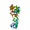 5es6SC 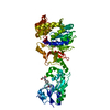 5es7C 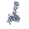 5es8C C: citing same article ( S: Starting model for refinement |
|---|---|
| Similar structure data |


-Links
 PDBj
PDBj


- Assembly
| Deposited unit | 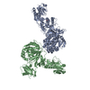
| ||||||||
|---|---|---|---|---|---|---|---|---|---|
| 1 | 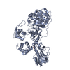
| ||||||||
| 2 | 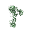
| ||||||||
| Unit cell |
|
-Components
| #1: Protein | Mass: 88063.180 Da / Num. of mol.: 2 / Fragment: UNP residues 2-766 Source method: isolated from a genetically manipulated source Source: (gene. exp.) Brevibacillus parabrevis (bacteria) / Gene: lgrA / Production host: #2: Chemical | ChemComp-PNS / | 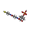  Mass: 358.348 Da / Num. of mol.: 1 / Source method: obtained synthetically / Formula: C11H23N2O7PS Mass: 358.348 Da / Num. of mol.: 1 / Source method: obtained synthetically / Formula: C11H23N2O7PS |
|---|
-Experimental details
-Experiment
| Experiment | Method: X-RAY DIFFRACTION / Number of used crystals: 1 |
|---|
- Sample preparation
| Crystal | Density Matthews: 4.51 Å3/Da / Density % sol: 72.71 % |
|---|---|
| Crystal grow | Temperature: 298 K / Method: vapor diffusion, sitting drop / Details: 1 M AmSO4, 0.1 M bis-Tris pH 5.5, 3% PEG 3350 |
-Data collection
| Diffraction | Mean temperature: 200 K | ||||||||||||||||||||||||||||||||||||||||||||||||||||||||||||||||||||||||||||||||||||||||||||||||||||||||||||||||||||||||||||||
|---|---|---|---|---|---|---|---|---|---|---|---|---|---|---|---|---|---|---|---|---|---|---|---|---|---|---|---|---|---|---|---|---|---|---|---|---|---|---|---|---|---|---|---|---|---|---|---|---|---|---|---|---|---|---|---|---|---|---|---|---|---|---|---|---|---|---|---|---|---|---|---|---|---|---|---|---|---|---|---|---|---|---|---|---|---|---|---|---|---|---|---|---|---|---|---|---|---|---|---|---|---|---|---|---|---|---|---|---|---|---|---|---|---|---|---|---|---|---|---|---|---|---|---|---|---|---|---|
| Diffraction source | Source: SYNCHROTRON / Site: CLSI / Beamline: 08ID-1 / Wavelength: 0.979 Å | ||||||||||||||||||||||||||||||||||||||||||||||||||||||||||||||||||||||||||||||||||||||||||||||||||||||||||||||||||||||||||||||
| Detector | Type: RAYONIX MX-300 / Detector: CCD / Date: Jan 21, 2015 | ||||||||||||||||||||||||||||||||||||||||||||||||||||||||||||||||||||||||||||||||||||||||||||||||||||||||||||||||||||||||||||||
| Radiation | Protocol: SINGLE WAVELENGTH / Monochromatic (M) / Laue (L): M / Scattering type: x-ray | ||||||||||||||||||||||||||||||||||||||||||||||||||||||||||||||||||||||||||||||||||||||||||||||||||||||||||||||||||||||||||||||
| Radiation wavelength | Wavelength: 0.979 Å / Relative weight: 1 | ||||||||||||||||||||||||||||||||||||||||||||||||||||||||||||||||||||||||||||||||||||||||||||||||||||||||||||||||||||||||||||||
| Reflection | Resolution: 3.77→48.45 Å / Num. obs: 32124 / % possible obs: 100 % / Redundancy: 11.2 % / Biso Wilson estimate: 190.98 Å2 / Rmerge(I) obs: 0.076 / Χ2: 1.041 / Net I/av σ(I): 27.219 / Net I/σ(I): 7.3 / Num. measured all: 327215 | ||||||||||||||||||||||||||||||||||||||||||||||||||||||||||||||||||||||||||||||||||||||||||||||||||||||||||||||||||||||||||||||
| Reflection shell | Diffraction-ID: 1 / Rejects: _
|
- Processing
| Software |
| ||||||||||||||||||||||||||||||||||||||||||||||||||||||||||||||||||||||||||||||
|---|---|---|---|---|---|---|---|---|---|---|---|---|---|---|---|---|---|---|---|---|---|---|---|---|---|---|---|---|---|---|---|---|---|---|---|---|---|---|---|---|---|---|---|---|---|---|---|---|---|---|---|---|---|---|---|---|---|---|---|---|---|---|---|---|---|---|---|---|---|---|---|---|---|---|---|---|---|---|---|
| Refinement | Method to determine structure: MOLECULAR REPLACEMENT Starting model: 5ES6 Resolution: 3.77→48.45 Å / SU ML: 0.7 / Cross valid method: FREE R-VALUE / σ(F): 1.34 / Phase error: 45.12 / Stereochemistry target values: ML
| ||||||||||||||||||||||||||||||||||||||||||||||||||||||||||||||||||||||||||||||
| Solvent computation | Shrinkage radii: 0.9 Å / VDW probe radii: 1.11 Å / Solvent model: FLAT BULK SOLVENT MODEL | ||||||||||||||||||||||||||||||||||||||||||||||||||||||||||||||||||||||||||||||
| Displacement parameters | Biso max: 541.5 Å2 / Biso mean: 261.0343 Å2 / Biso min: 160.85 Å2 | ||||||||||||||||||||||||||||||||||||||||||||||||||||||||||||||||||||||||||||||
| Refinement step | Cycle: final / Resolution: 3.77→48.45 Å
| ||||||||||||||||||||||||||||||||||||||||||||||||||||||||||||||||||||||||||||||
| Refine LS restraints |
| ||||||||||||||||||||||||||||||||||||||||||||||||||||||||||||||||||||||||||||||
| LS refinement shell | Refine-ID: X-RAY DIFFRACTION / Total num. of bins used: 12 / % reflection obs: 100 %
|
