 Movie
Movie Controller

[English] 日本語
 Yorodumi
- PDB-6wty: Plasmodium vivax reticulocyte binding protein 2b (PvRBP2b) bound ... -
+ Open data

- Basic information
| Entry | Database: PDB / ID: 6wty | |||||||||||||||
|---|---|---|---|---|---|---|---|---|---|---|---|---|---|---|---|---|
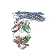
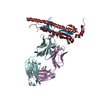
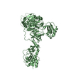
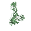
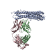
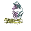
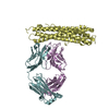
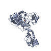
| Title | Plasmodium vivax reticulocyte binding protein 2b (PvRBP2b) bound to human monoclonal antibody 253245 | |||||||||||||||
 Components Components |
| |||||||||||||||
 Keywords Keywords | CELL INVASION / invasion / plasmodium vivax / malaria / antibody complex | |||||||||||||||
| Function / homology | NBD94 domain / Nucleotide-Binding Domain 94 of RH / Immunoglobulins / Immunoglobulin-like / Sandwich / Mainly Beta / Reticulocyte-binding protein 2b Function and homology information Function and homology information | |||||||||||||||
| Biological species |   Homo sapiens (human) Homo sapiens (human) | |||||||||||||||
| Method |  X-RAY DIFFRACTION / SYNCHROTRON / MOLECULAR REPLACEMENT / Resolution: 3.481 Å X-RAY DIFFRACTION / SYNCHROTRON / MOLECULAR REPLACEMENT / Resolution: 3.481 Å | |||||||||||||||
 Authors Authors | Chan, L.J. / Dietrich, M.H. / Tham, W.H. | |||||||||||||||
| Funding support |  Australia, 4items Australia, 4items
| |||||||||||||||
 Citation Citation | Journal: Nat Commun / Year: 2021 Title: Naturally acquired blocking human monoclonal antibodies to Plasmodium vivax reticulocyte binding protein 2b. Authors: Chan, L.J. / Gandhirajan, A. / Carias, L.L. / Dietrich, M.H. / Vadas, O. / Visentin, R. / Franca, C.T. / Menant, S. / Soldati-Favre, D. / Mueller, I. / King, C.L. / Tham, W.H. | |||||||||||||||
| History |
|
- Structure visualization
| Structure viewer | Molecule: MolmilJmol/JSmol |
|---|
- Downloads & links
-Download
| PDBx/mmCIF format | 6wty.cif.gz | 519.8 KB | Display | PDBx/mmCIF format |
|---|---|---|---|---|
| PDB format | pdb6wty.ent.gz | 407.3 KB | Display | PDB format |
| PDBx/mmJSON format | 6wty.json.gz | Tree view | PDBx/mmJSON format | |
| Others |  Other downloads Other downloads |
-Validation report
| Arichive directory | https://data.pdbj.org/pub/pdb/validation_reports/wt/6wtyftp://data.pdbj.org/pub/pdb/validation_reports/wt/6wty | HTTPS FTP |
|---|
-Related structure data
| Related structure data |  6wm9C  6wn1C  6wnoC  6wozC  6wqoC  6wtuC  6wtvC  5w53S C: citing same article ( S: Starting model for refinement |
|---|---|
| Similar structure data |
-Links
 PDBj
PDBj

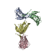
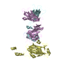
- Assembly
| Deposited unit | 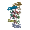
| ||||||||
|---|---|---|---|---|---|---|---|---|---|
| 1 | 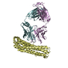
| ||||||||
| 2 | 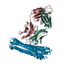
| ||||||||
| 3 | 
| ||||||||
| 4 | 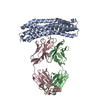
| ||||||||
| Unit cell |
|
-Components
| #1: Protein | Mass: 36519.789 Da / Num. of mol.: 4 Source method: isolated from a genetically manipulated source Source: (gene. exp.) Strain: Salvador I / Gene: PVX_094255 / Plasmid: pET32a(+) / Production host:  #2: Antibody | Mass: 23470.975 Da / Num. of mol.: 4 Source method: isolated from a genetically manipulated source Source: (gene. exp.) Homo sapiens (human) / Cell line (production host): Expi293 / Production host: Homo sapiens (human)#3: Antibody | Mass: 25668.795 Da / Num. of mol.: 4 Source method: isolated from a genetically manipulated source Source: (gene. exp.) Homo sapiens (human) / Cell line (production host): Expi293 / Production host: Homo sapiens (human)Has protein modification | Y | |
|---|
-Experimental details
-Experiment
| Experiment | Method: X-RAY DIFFRACTION / Number of used crystals: 1 |
|---|
- Sample preparation
| Crystal | Density Matthews: 2.46 Å3/Da / Density % sol: 49.95 % |
|---|---|
| Crystal grow | Temperature: 295 K / Method: vapor diffusion, hanging drop / pH: 4 Details: 0.2 M sodium dihydrogen phosphate pH 4.0, 16% (w/v) PEG 3,350 |
-Data collection
| Diffraction | Mean temperature: 100 K / Serial crystal experiment: N | ||||||||||||||||||||||||||||||||||||||||||||||||||||||||||||||||||||||||||||||||||||||||||||||||||||
|---|---|---|---|---|---|---|---|---|---|---|---|---|---|---|---|---|---|---|---|---|---|---|---|---|---|---|---|---|---|---|---|---|---|---|---|---|---|---|---|---|---|---|---|---|---|---|---|---|---|---|---|---|---|---|---|---|---|---|---|---|---|---|---|---|---|---|---|---|---|---|---|---|---|---|---|---|---|---|---|---|---|---|---|---|---|---|---|---|---|---|---|---|---|---|---|---|---|---|---|---|---|
| Diffraction source | Source: SYNCHROTRON / Site: Australian Synchrotron / Beamline: MX2 / Wavelength: 0.9537 Å | ||||||||||||||||||||||||||||||||||||||||||||||||||||||||||||||||||||||||||||||||||||||||||||||||||||
| Detector | Type: DECTRIS EIGER X 16M / Detector: PIXEL / Date: Sep 29, 2018 | ||||||||||||||||||||||||||||||||||||||||||||||||||||||||||||||||||||||||||||||||||||||||||||||||||||
| Radiation | Protocol: SINGLE WAVELENGTH / Monochromatic (M) / Laue (L): M / Scattering type: x-ray | ||||||||||||||||||||||||||||||||||||||||||||||||||||||||||||||||||||||||||||||||||||||||||||||||||||
| Radiation wavelength | Wavelength: 0.9537 Å / Relative weight: 1 | ||||||||||||||||||||||||||||||||||||||||||||||||||||||||||||||||||||||||||||||||||||||||||||||||||||
| Reflection | Resolution: 3.48→48.795 Å / Num. obs: 43497 / % possible obs: 98.7 % / Redundancy: 5.388 % / Biso Wilson estimate: 60.59 Å2 / CC1/2: 0.85 / Rmerge(I) obs: 0.772 / Rrim(I) all: 0.857 / Χ2: 0.613 / Net I/σ(I): 2.54 / Num. measured all: 234345 | ||||||||||||||||||||||||||||||||||||||||||||||||||||||||||||||||||||||||||||||||||||||||||||||||||||
| Reflection shell | Diffraction-ID: 1
|
- Processing
| Software |
| |||||||||||||||||||||||||||||||||||||||||||||||||||||||||||||||||||||||||||||||||||||||||||||||||||||||||
|---|---|---|---|---|---|---|---|---|---|---|---|---|---|---|---|---|---|---|---|---|---|---|---|---|---|---|---|---|---|---|---|---|---|---|---|---|---|---|---|---|---|---|---|---|---|---|---|---|---|---|---|---|---|---|---|---|---|---|---|---|---|---|---|---|---|---|---|---|---|---|---|---|---|---|---|---|---|---|---|---|---|---|---|---|---|---|---|---|---|---|---|---|---|---|---|---|---|---|---|---|---|---|---|---|---|---|
| Refinement | Method to determine structure: MOLECULAR REPLACEMENT Starting model: 5W53 Resolution: 3.481→48.701 Å / Cross valid method: THROUGHOUT / σ(F): 1498.83 / Phase error: 42.24 / Stereochemistry target values: TWIN_LSQ_F Details: Pseudo-merohedral twinning was detected. Therefore the structure was refined in the presence of the twinning operator (h, -k, -h-l) yielding an associated twin fraction of 0.15.
| |||||||||||||||||||||||||||||||||||||||||||||||||||||||||||||||||||||||||||||||||||||||||||||||||||||||||
| Solvent computation | Shrinkage radii: 0.9 Å / VDW probe radii: 1.11 Å / Solvent model: FLAT BULK SOLVENT MODEL | |||||||||||||||||||||||||||||||||||||||||||||||||||||||||||||||||||||||||||||||||||||||||||||||||||||||||
| Displacement parameters | Biso max: 153.14 Å2 / Biso mean: 74.7432 Å2 / Biso min: 32.68 Å2 | |||||||||||||||||||||||||||||||||||||||||||||||||||||||||||||||||||||||||||||||||||||||||||||||||||||||||
| Refinement step | Cycle: final / Resolution: 3.481→48.701 Å
| |||||||||||||||||||||||||||||||||||||||||||||||||||||||||||||||||||||||||||||||||||||||||||||||||||||||||
| LS refinement shell | Refine-ID: X-RAY DIFFRACTION / Rfactor Rfree error: 0 / Total num. of bins used: 14
|
