| Entry | Database: PDB / ID: 5w53
|
|---|
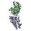
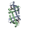
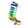
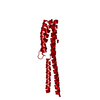
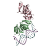
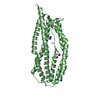
| Title | Crystal structure of the erythrocyte-binding domain from Plasmodium vivax reticulocyte-binding protein 2b (PvRBP2b) |
|---|
 Components Components | Reticulocyte binding protein 2, putative |
|---|
 Keywords Keywords | CELL INVASION / reticulocyte-binding / alpha-helical |
|---|
| Function / homology | NBD94 domain / Nucleotide-Binding Domain 94 of RH / : / THIOCYANATE ION / Reticulocyte-binding protein 2b Function and homology information Function and homology information |
|---|
| Biological species |   Plasmodium vivax (malaria parasite P. vivax) Plasmodium vivax (malaria parasite P. vivax) |
|---|
| Method |  X-RAY DIFFRACTION / SYNCHROTRON / MOLECULAR REPLACEMENT / Resolution: 1.71 Å X-RAY DIFFRACTION / SYNCHROTRON / MOLECULAR REPLACEMENT / Resolution: 1.71 Å |
|---|
 Authors Authors | Gruszczyk, J. / Tham, W.H. |
|---|
 Citation Citation | Journal: Science / Year: 2018
Title: Transferrin receptor 1 is a reticulocyte-specific receptor for Plasmodium vivax.
Authors: Gruszczyk, J. / Kanjee, U. / Chan, L.J. / Menant, S. / Malleret, B. / Lim, N.T.Y. / Schmidt, C.Q. / Mok, Y.F. / Lin, K.M. / Pearson, R.D. / Rangel, G. / Smith, B.J. / Call, M.J. / Weekes, M. ...Authors: Gruszczyk, J. / Kanjee, U. / Chan, L.J. / Menant, S. / Malleret, B. / Lim, N.T.Y. / Schmidt, C.Q. / Mok, Y.F. / Lin, K.M. / Pearson, R.D. / Rangel, G. / Smith, B.J. / Call, M.J. / Weekes, M.P. / Griffin, M.D.W. / Murphy, J.M. / Abraham, J. / Sriprawat, K. / Menezes, M.J. / Ferreira, M.U. / Russell, B. / Renia, L. / Duraisingh, M.T. / Tham, W.H. |
|---|
| History | | Deposition | Jun 13, 2017 | Deposition site: RCSB / Processing site: RCSB |
|---|
| Revision 1.0 | Nov 29, 2017 | Provider: repository / Type: Initial release |
|---|
| Revision 1.1 | Jan 17, 2018 | Group: Database references / Category: citation / citation_author
Item: _citation.journal_volume / _citation.page_first ..._citation.journal_volume / _citation.page_first / _citation.page_last / _citation.pdbx_database_id_DOI / _citation.pdbx_database_id_PubMed / _citation.year |
|---|
| Revision 1.2 | Oct 4, 2023 | Group: Data collection / Database references ...Data collection / Database references / Derived calculations / Refinement description
Category: chem_comp_atom / chem_comp_bond ...chem_comp_atom / chem_comp_bond / database_2 / pdbx_initial_refinement_model / pdbx_struct_conn_angle / struct_conn
Item: _database_2.pdbx_DOI / _database_2.pdbx_database_accession ..._database_2.pdbx_DOI / _database_2.pdbx_database_accession / _pdbx_struct_conn_angle.ptnr1_auth_asym_id / _pdbx_struct_conn_angle.ptnr1_auth_comp_id / _pdbx_struct_conn_angle.ptnr1_auth_seq_id / _pdbx_struct_conn_angle.ptnr1_label_asym_id / _pdbx_struct_conn_angle.ptnr1_label_atom_id / _pdbx_struct_conn_angle.ptnr1_label_comp_id / _pdbx_struct_conn_angle.ptnr1_label_seq_id / _pdbx_struct_conn_angle.ptnr1_symmetry / _pdbx_struct_conn_angle.ptnr2_auth_asym_id / _pdbx_struct_conn_angle.ptnr2_auth_seq_id / _pdbx_struct_conn_angle.ptnr2_label_asym_id / _pdbx_struct_conn_angle.ptnr2_symmetry / _pdbx_struct_conn_angle.ptnr3_auth_asym_id / _pdbx_struct_conn_angle.ptnr3_auth_comp_id / _pdbx_struct_conn_angle.ptnr3_auth_seq_id / _pdbx_struct_conn_angle.ptnr3_label_asym_id / _pdbx_struct_conn_angle.ptnr3_label_comp_id / _pdbx_struct_conn_angle.ptnr3_label_seq_id / _pdbx_struct_conn_angle.ptnr3_symmetry / _pdbx_struct_conn_angle.value / _struct_conn.pdbx_dist_value / _struct_conn.ptnr1_auth_asym_id / _struct_conn.ptnr1_auth_comp_id / _struct_conn.ptnr1_auth_seq_id / _struct_conn.ptnr1_label_asym_id / _struct_conn.ptnr1_label_atom_id / _struct_conn.ptnr1_label_comp_id / _struct_conn.ptnr1_label_seq_id / _struct_conn.ptnr1_symmetry / _struct_conn.ptnr2_auth_asym_id / _struct_conn.ptnr2_auth_comp_id / _struct_conn.ptnr2_auth_seq_id / _struct_conn.ptnr2_label_asym_id / _struct_conn.ptnr2_label_atom_id / _struct_conn.ptnr2_label_comp_id / _struct_conn.ptnr2_label_seq_id / _struct_conn.ptnr2_symmetry |
|---|
| Revision 1.3 | Nov 13, 2024 | Group: Structure summary / Category: pdbx_entry_details / pdbx_modification_feature |
|---|
|
|---|
 Movie
Movie Controller

 Yorodumi
 Open data
 Basic information
 Structure visualization
 Downloads & links
 Other downloads


 PDBj
PDBj
 Assembly
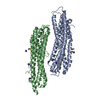


 Mass: 58.082 Da / Num. of mol.: 5 / Source method: obtained synthetically / Formula: CNS
 Mass: 39.098 Da / Num. of mol.: 8 / Source method: obtained synthetically / Formula: K
 Mass: 18.015 Da / Num. of mol.: 824 / Source method: isolated from a natural source / Formula: H2O
 Sample preparation
 / Beamline: MX2 / Wavelength: 0.9537 Å
 Processing