Mass: 65.409 Da / Num. of mol.: 2 / Source method: obtained synthetically / Formula: Zn
Has protein modification
N
Sequence details
THE AUTHORS CRYSTALLIZED THE FRAGMENT PROTEIN, RESIDUES 390-1365 FOR CHAIN A. THE SEQUENCES OF ...THE AUTHORS CRYSTALLIZED THE FRAGMENT PROTEIN, RESIDUES 390-1365 FOR CHAIN A. THE SEQUENCES OF RESIDUES 505-521 AND 712-849 (THE UNK PARTS) ARE AS FOLLOWS. (505)AKAARPPWEPPKTKLDEDLESSSESECESDEDSTCSSSSDSEVFDVIAEIKRKKAHPDRLHDE(521) AND (735)ALVPEEEIANMLQWEELEWQKYAEECKGMIVTNPGTKPSSVRIDQLDREQFNPDVITFPIIVHFGIRPAQLSYAGDPQYQKLWKSYVKLRHLLANSPKVKQTDKQKLAQREEALQKIRQKNTMRREVTVELSSQGFWK(849) THE AUTHORS COULD OBSERVE THREE PARTS (505-520,735-745,749-756). BUT THEY ARE NOT SURE WHICH PART CORRESPONDS TO THESE OBSERVED RESIDUES. SO THE RESIDUE NUMBERS OF UNK ARE MEANINGLESS.
-
Experimental details
-
Experiment
Experiment
Method: X-RAY DIFFRACTION / Number of used crystals: 1
-
Sample preparation
Crystal
Density Matthews: 3.34 Å3/Da / Density % sol: 63.17 %
Monochromator: DCM Si (111) Crystal / Protocol: SINGLE WAVELENGTH / Monochromatic (M) / Laue (L): M / Scattering type: x-ray
Radiation wavelength
Wavelength: 1 Å / Relative weight: 1
Reflection
Redundancy: 7.5 % / Number: 183042 / Rmerge(I) obs: 0.061 / Χ2: 0.93 / D res high: 3.3 Å / D res low: 20 Å / Num. obs: 24319 / % possible obs: 99.6
Diffraction reflection shell
Highest resolution (Å)
Lowest resolution (Å)
ID
Rmerge(I) obs
Chi squared
Redundancy
8.72
20
1
0.029
0.71
7.2
7.02
8.72
1
0.031
0.719
7.6
6.16
7.02
1
0.038
0.733
7.2
5.61
6.16
1
0.044
0.751
7.3
5.22
5.61
1
0.051
0.822
7.4
4.91
5.22
1
0.058
0.958
7.5
4.67
4.91
1
0.066
1.131
7.5
4.47
4.67
1
0.07
1.097
7.6
4.3
4.47
1
0.08
1.004
7.6
4.15
4.3
1
0.092
0.973
7.6
4.02
4.15
1
0.109
0.913
7.6
3.91
4.02
1
0.134
0.932
7.6
3.81
3.91
1
0.165
0.958
7.6
3.71
3.81
1
0.198
0.938
7.6
3.63
3.71
1
0.219
1
7.6
3.55
3.63
1
0.328
0.969
7.6
3.48
3.55
1
0.357
0.987
7.6
3.42
3.48
1
0.436
0.978
7.6
3.36
3.42
1
0.561
1.031
7.6
3.3
3.36
1
0.65
0.97
7.6
Reflection
Resolution: 3.2→20 Å / Num. obs: 26854 / % possible obs: 99.9 % / Redundancy: 7.3 % / Biso Wilson estimate: 49.52 Å2 / Rmerge(I) obs: 0.059 / Net I/σ(I): 10.7
Reflection shell
Resolution: 3.2→3.25 Å / Redundancy: 7.1 % / Rmerge(I) obs: 0.577 / % possible all: 100
-
Processing
Software
Name
Version
Classification
PHENIX
1.9_1692
refinement
SCALEPACK
datascaling
PDB_EXTRACT
3.15
dataextraction
HKL-2000
datareduction
PHENIX
phasing
Refinement
Method to determine structure: MOLECULAR REPLACEMENT / Resolution: 3.2→19.93 Å / Cross valid method: FREE R-VALUE / Details: ANOMALOUS DATA WAS USED FOR REFINEMENT.
Rfactor
Num. reflection
% reflection
Rfree
0.3
3143
8.86 %
Rwork
0.267
-
-
obs
-
22695
67.3 %
Displacement parameters
Biso mean: 61.56 Å2
Refinement step
Cycle: LAST / Resolution: 3.2→19.93 Å
Protein
Nucleic acid
Ligand
Solvent
Total
Num. atoms
6157
0
2
0
6159
LS refinement shell
Resolution: 3.2→3.25 Å
Rfactor
Num. reflection
% reflection
Rfree
0.5292
49
-
Rwork
0.3999
504
-
obs
-
-
24 %
Refinement TLS params.
Method: refined / Refine-ID: X-RAY DIFFRACTION
ID
L11 (°2)
L12 (°2)
L13 (°2)
L22 (°2)
L23 (°2)
L33 (°2)
S11 (Å °)
S12 (Å °)
S13 (Å °)
S21 (Å °)
S22 (Å °)
S23 (Å °)
S31 (Å °)
S32 (Å °)
S33 (Å °)
T11 (Å2)
T12 (Å2)
T13 (Å2)
T22 (Å2)
T23 (Å2)
T33 (Å2)
Origin x (Å)
Origin y (Å)
Origin z (Å)
1
0.1424
0.1956
-0.1697
0.4613
0.0353
0.4932
0.0845
-0.3172
-0.0074
0.1706
-0.0585
0.411
-0.1365
-0.4497
-0.1404
0.1013
0.1409
0.3104
0.7174
0.2228
0.0968
116.7057
-7.295
165.7279
2
0.1777
0.0605
0.0129
0.6184
0.078
0.3703
0.0315
0.0704
-0.1519
-0.698
0.2601
0.0935
0.072
-0.2918
0.3676
0.6151
-0.4712
-0.4146
-0.1461
0.146
0.4659
114.1126
-22.8182
118.5785
Refinement TLS group
ID
Refine-ID
Refine TLS-ID
Selection details
1
X-RAY DIFFRACTION
1
(chain 'A' and ((resseq411:711) or (resseq735:756) or (resseq850:863) or (resseq904:929) or (resseq1401;1402)))
2
X-RAY DIFFRACTION
2
(chain 'A' and ((resseq864:903) or (resseq958:1333))) or (chain 'B') or (chain 'C')
+
About Yorodumi
-
News
-
Feb 9, 2022. New format data for meta-information of EMDB entries
New format data for meta-information of EMDB entries
Version 3 of the EMDB header file is now the official format.
The previous official version 1.9 will be removed from the archive.
In the structure databanks used in Yorodumi, some data are registered as the other names, "COVID-19 virus" and "2019-nCoV". Here are the details of the virus and the list of structure data.
Jan 31, 2019. EMDB accession codes are about to change! (news from PDBe EMDB page)
EMDB accession codes are about to change! (news from PDBe EMDB page)
The allocation of 4 digits for EMDB accession codes will soon come to an end. Whilst these codes will remain in use, new EMDB accession codes will include an additional digit and will expand incrementally as the available range of codes is exhausted. The current 4-digit format prefixed with “EMD-” (i.e. EMD-XXXX) will advance to a 5-digit format (i.e. EMD-XXXXX), and so on. It is currently estimated that the 4-digit codes will be depleted around Spring 2019, at which point the 5-digit format will come into force.
The EM Navigator/Yorodumi systems omit the EMD- prefix.
Related info.:Q: What is EMD? / ID/Accession-code notation in Yorodumi/EM Navigator
Yorodumi is a browser for structure data from EMDB, PDB, SASBDB, etc.
This page is also the successor to EM Navigator detail page, and also detail information page/front-end page for Omokage search.
The word "yorodu" (or yorozu) is an old Japanese word meaning "ten thousand". "mi" (miru) is to see.
Related info.:EMDB / PDB / SASBDB / Comparison of 3 databanks / Yorodumi Search / Aug 31, 2016. New EM Navigator & Yorodumi / Yorodumi Papers / Jmol/JSmol / Function and homology information / Changes in new EM Navigator and Yorodumi
 Movie
Movie Controller

 Yorodumi
 Open data
 Basic information
 Components
 Keywords
 Function and homology information
 Homo sapiens (human)
 X-RAY DIFFRACTION /
  Authors
 Korea, Republic Of, 1items
  Citation
 Structure visualization
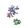
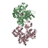
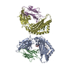
 Downloads & links
 Other downloads


 PDBj
PDBj


 Assembly
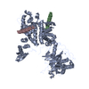

 Mass: 65.409 Da / Num. of mol.: 2 / Source method: obtained synthetically / Formula: Zn
 Sample preparation
 Processing