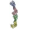
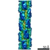
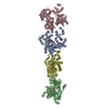
CONTRACTILE PROTEIN / ARCHAEA / CRENARCHAEOTA / CYTOSKELETON / EVOLUTION
Function / homology
Function and homology information
Hydrolases; Acting on acid anhydrides; Acting on acid anhydrides to facilitate cellular and subcellular movement / cytoskeleton / hydrolase activity / ATP binding Similarity search - Function
Protocol: SINGLE WAVELENGTH / Monochromatic (M) / Laue (L): M / Scattering type: x-ray
Radiation wavelength
Wavelength: 0.9793 Å / Relative weight: 1
Reflection
Resolution: 3.34→210.8 Å / Num. obs: 40652 / % possible obs: 98 % / Observed criterion σ(I): 2.5 / Redundancy: 4.2 % / Rmerge(I) obs: 0.08 / Net I/σ(I): 12
Reflection shell
Resolution: 3.34→3.42 Å / Rmerge(I) obs: 0.3 / Mean I/σ(I) obs: 2.5 / % possible all: 82
-
Processing
Software
Name
Version
Classification
REFMAC
5.6.0117
refinement
HKL-3000
datareduction
HKL-3000
datascaling
HKL-3000
phasing
Refinement
Method to determine structure: SAD Starting model: NONE Resolution: 3.34→210.79 Å / Cor.coef. Fo:Fc: 0.937 / Cor.coef. Fo:Fc free: 0.909 / SU B: 55.156 / SU ML: 0.397 / Cross valid method: THROUGHOUT / ESU R Free: 0.504 / Stereochemistry target values: MAXIMUM LIKELIHOOD / Details: HYDROGENS HAVE BEEN ADDED IN THE RIDING POSITIONS.
Rfactor
Num. reflection
% reflection
Selection details
Rfree
0.24781
2033
5 %
RANDOM
Rwork
0.20723
-
-
-
obs
0.20929
38545
98.25 %
-
Solvent computation
Ion probe radii: 0.8 Å / Shrinkage radii: 0.8 Å / VDW probe radii: 1.2 Å / Solvent model: MASK
 Movie
Movie Controller


 Open data
 Basic information
 Components
 Keywords
 Function and homology information
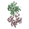
 PYROBACULUM CALIDIFONTIS (archaea)
 X-RAY DIFFRACTION /
  Authors
 Citation
 Structure visualization
 Downloads & links
 Other downloads


 PDBj
PDBj


 Assembly
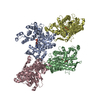


 Mass: 427.201 Da / Num. of mol.: 3 / Source method: obtained synthetically / Formula: C10H15N5O10P2 / Comment: ADP, energy-carrying molecule*YM
 Mass: 24.305 Da / Num. of mol.: 1 / Source method: obtained synthetically / Formula: Mg
 Sample preparation
 / Beamline: ID29 / Wavelength: 0.9793
  Processing