 Movie
Movie Controller

[English] 日本語
 Yorodumi
- PDB-1m6n: Crystal structure of the SecA translocation ATPase from Bacillus ... -
+ Open data

- Basic information
| Entry | Database: PDB / ID: 1m6n | ||||||
|---|---|---|---|---|---|---|---|
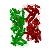
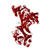
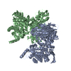
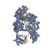
| Title | Crystal structure of the SecA translocation ATPase from Bacillus subtilis | ||||||
 Components Components | Preprotein translocase secA | ||||||
 Keywords Keywords | PROTEIN TRANSPORT / protein translocation / atpase / transmembrane transport / helicase family structure / mechanochemisty | ||||||
| Function / homology |  Function and homology information Function and homology informationcell envelope Sec protein transport complex / protein-exporting ATPase activity / protein-secreting ATPase / protein transport by the Sec complex / intracellular protein transmembrane transport / protein import / protein targeting / membrane raft / ATP binding / metal ion binding ...cell envelope Sec protein transport complex / protein-exporting ATPase activity / protein-secreting ATPase / protein transport by the Sec complex / intracellular protein transmembrane transport / protein import / protein targeting / membrane raft / ATP binding / metal ion binding / plasma membrane / cytoplasm Similarity search - Function | ||||||
| Biological species |  | ||||||
| Method |  X-RAY DIFFRACTION / SYNCHROTRON / MIR / Resolution: 2.7 Å X-RAY DIFFRACTION / SYNCHROTRON / MIR / Resolution: 2.7 Å | ||||||
 Authors Authors | Hunt, J.F. / Weinkauf, S. / Henry, L. / Fak, J.J. / McNicholas, P. / Oliver, D.B. / Deisenhofer, J. | ||||||
 Citation Citation | Journal: Science / Year: 2002 Title: Nucleotide Control of Interdomain Interactions in the Conformational Reaction Cycle of SecA Authors: Hunt, J.F. / Weinkauf, S. / Henry, L. / Fak, J.J. / McNicholas, P. / Oliver, D.B. / Deisenhofer, J. #1: Journal: To be Published / Year: 2002Title: Ping-pong cross-validation in real space: a method to increase the phasing power of a partial model without risk of phase bias Authors: Hunt, J.F. / Deisenhofer, J. #2: Journal: Acta Crystallogr.,Sect.D / Year: 2001Title: Conformational stabilization and crystallization of the SecA translocation ATPase from Bacillus subtilis Authors: Weinkauf, S. / Hunt, J.F. / Scheuring, J. / Henry, L. / Fak, J.J. / Oliver, D.B. / Deisenhofer, J. | ||||||
| History |
|
- Structure visualization
| Structure viewer | Molecule: MolmilJmol/JSmol |
|---|
- Downloads & links
-Download
| PDBx/mmCIF format | 1m6n.cif.gz | 172 KB | Display | PDBx/mmCIF format |
|---|---|---|---|---|
| PDB format | pdb1m6n.ent.gz | 136.9 KB | Display | PDB format |
| PDBx/mmJSON format | 1m6n.json.gz | Tree view | PDBx/mmJSON format | |
| Others |  Other downloads Other downloads |
-Validation report
| Arichive directory | https://data.pdbj.org/pub/pdb/validation_reports/m6/1m6nftp://data.pdbj.org/pub/pdb/validation_reports/m6/1m6n | HTTPS FTP |
|---|
-Related structure data


-Links
 PDBj
PDBj- Assembly
| Deposited unit | 
| ||||||||||
|---|---|---|---|---|---|---|---|---|---|---|---|
| 1 | 
| ||||||||||
| Unit cell |
| ||||||||||
| Details | The other subunit in the physiological dimer is generated by 1-y, 1-x, 2/3-z (4_665) |
-Components
| #1: Protein | Mass: 91393.758 Da / Num. of mol.: 1 Source method: isolated from a genetically manipulated source Source: (gene. exp.) | ||
|---|---|---|---|
| #2: Chemical | ChemComp-SO4 /   Mass: 96.063 Da / Num. of mol.: 7 / Source method: obtained synthetically / Formula: SO4 Mass: 96.063 Da / Num. of mol.: 7 / Source method: obtained synthetically / Formula: SO4#3: Water | ChemComp-HOH / |  Mass: 18.015 Da / Num. of mol.: 45 / Source method: isolated from a natural source / Formula: H2O Mass: 18.015 Da / Num. of mol.: 45 / Source method: isolated from a natural source / Formula: H2O |
-Experimental details
-Experiment
| Experiment | Method: X-RAY DIFFRACTION / Number of used crystals: 1 |
|---|
- Sample preparation
| Crystal | Density Matthews: 4.06 Å3/Da / Density % sol: 69.73 % | ||||||||||||||||||||||||||||||||||||||||||||||||
|---|---|---|---|---|---|---|---|---|---|---|---|---|---|---|---|---|---|---|---|---|---|---|---|---|---|---|---|---|---|---|---|---|---|---|---|---|---|---|---|---|---|---|---|---|---|---|---|---|---|
| Crystal grow | Temperature: 299 K / Method: vapor diffusion, hanging drop / pH: 7 Details: 2 M ammonium sulfate, 30% glycerol, 1 mM DTT, 100 mM BES, pH 7.0, VAPOR DIFFUSION, HANGING DROP at 299K | ||||||||||||||||||||||||||||||||||||||||||||||||
| Crystal grow | *PLUS Details: Weinkauf, S., (2001) Acta Crystallogr., Sect.D, 57, 559. | ||||||||||||||||||||||||||||||||||||||||||||||||
| Components of the solutions | *PLUS
|
-Data collection
| Diffraction | Mean temperature: 100 K |
|---|---|
| Diffraction source | Source: SYNCHROTRON / Site: ESRF  / Beamline: ID2 / Wavelength: 0.986 Å / Beamline: ID2 / Wavelength: 0.986 Å |
| Detector | Type: MARRESEARCH / Detector: AREA DETECTOR / Date: Dec 15, 1997 |
| Radiation | Protocol: SINGLE WAVELENGTH / Monochromatic (M) / Laue (L): M / Scattering type: x-ray |
| Radiation wavelength | Wavelength: 0.986 Å / Relative weight: 1 |
| Reflection | Resolution: 2.7→45 Å / Num. all: 40576 / Num. obs: 40372 / % possible obs: 99.5 % / Observed criterion σ(I): -3 / Redundancy: 6.9 % / Biso Wilson estimate: 62 Å2 / Rmerge(I) obs: 0.073 / Rsym value: 0.073 / Net I/σ(I): 18.9 |
| Reflection shell | Resolution: 2.7→2.75 Å / Mean I/σ(I) obs: 2.14 / Num. unique all: 2018 / % possible all: 0.973 |
| Reflection | *PLUS Highest resolution: 2.7 Å / Lowest resolution: 50 Å / % possible obs: 99.6 % / Rmerge(I) obs: 0.078 |
- Processing
| Software |
| ||||||||||||||||||||||||||||||||||||||||||||||||||||||||||||||||||||||||||||||||
|---|---|---|---|---|---|---|---|---|---|---|---|---|---|---|---|---|---|---|---|---|---|---|---|---|---|---|---|---|---|---|---|---|---|---|---|---|---|---|---|---|---|---|---|---|---|---|---|---|---|---|---|---|---|---|---|---|---|---|---|---|---|---|---|---|---|---|---|---|---|---|---|---|---|---|---|---|---|---|---|---|---|
| Refinement | Method to determine structure: MIR / Resolution: 2.7→45.24 Å / Rfactor Rfree error: 0.007 / Data cutoff high absF: 2936891.3 / Data cutoff low absF: 0 / Isotropic thermal model: RESTRAINED / Cross valid method: THROUGHOUT / σ(F): 2 / Stereochemistry target values: Engh & Huber
| ||||||||||||||||||||||||||||||||||||||||||||||||||||||||||||||||||||||||||||||||
| Solvent computation | Solvent model: FLAT MODEL / Bsol: 91.5959 Å2 / ksol: 0.330937 e/Å3 | ||||||||||||||||||||||||||||||||||||||||||||||||||||||||||||||||||||||||||||||||
| Displacement parameters | Biso mean: 98.4 Å2
| ||||||||||||||||||||||||||||||||||||||||||||||||||||||||||||||||||||||||||||||||
| Refine analyze |
| ||||||||||||||||||||||||||||||||||||||||||||||||||||||||||||||||||||||||||||||||
| Refinement step | Cycle: LAST / Resolution: 2.7→45.24 Å
| ||||||||||||||||||||||||||||||||||||||||||||||||||||||||||||||||||||||||||||||||
| Refine LS restraints |
| ||||||||||||||||||||||||||||||||||||||||||||||||||||||||||||||||||||||||||||||||
| LS refinement shell | Resolution: 2.7→2.87 Å / Rfactor Rfree error: 0.028 / Total num. of bins used: 6
| ||||||||||||||||||||||||||||||||||||||||||||||||||||||||||||||||||||||||||||||||
| Xplor file |
| ||||||||||||||||||||||||||||||||||||||||||||||||||||||||||||||||||||||||||||||||
| Refinement | *PLUS Rfactor obs: 0.22 / Rfactor Rfree: 0.304 / Rfactor Rwork: 0.221 | ||||||||||||||||||||||||||||||||||||||||||||||||||||||||||||||||||||||||||||||||
| Solvent computation | *PLUS | ||||||||||||||||||||||||||||||||||||||||||||||||||||||||||||||||||||||||||||||||
| Displacement parameters | *PLUS | ||||||||||||||||||||||||||||||||||||||||||||||||||||||||||||||||||||||||||||||||
| Refine LS restraints | *PLUS
| ||||||||||||||||||||||||||||||||||||||||||||||||||||||||||||||||||||||||||||||||
| LS refinement shell | *PLUS Rfactor Rfree: 0.381 / Rfactor Rwork: 0.355 |
