 Movie
Movie Controller

[English] 日本語
 Yorodumi
- PDB-5xao: Crystal structure of Phaeospaeria nodrum fructosyl peptide oxidas... -
+ Open data

- Basic information
| Entry | Database: PDB / ID: 5xao | ||||||
|---|---|---|---|---|---|---|---|
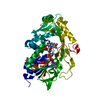
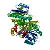
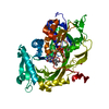
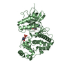
| Title | Crystal structure of Phaeospaeria nodrum fructosyl peptide oxidase mutant Asn56Ala in complexes with sodium and chloride ions | ||||||
 Components Components | Uncharacterized protein | ||||||
 Keywords Keywords | OXIDOREDUCTASE / Fructosyl peptide oxidase / FAD | ||||||
| Function / homology |  Function and homology information Function and homology informationsaccharopine oxidase activity / sarcosine oxidase activity / flavin adenine dinucleotide binding Similarity search - Function | ||||||
| Biological species |  Phaeosphaeria nodorum (fungus) Phaeosphaeria nodorum (fungus) | ||||||
| Method |  X-RAY DIFFRACTION / MOLECULAR REPLACEMENT / Resolution: 1.8 Å X-RAY DIFFRACTION / MOLECULAR REPLACEMENT / Resolution: 1.8 Å | ||||||
 Authors Authors | Yoshida, H. / Kamitori, S. / Sode, K. | ||||||
 Citation Citation | Journal: Sci Rep / Year: 2017 Title: X-ray structures of fructosyl peptide oxidases revealing residues responsible for gating oxygen access in the oxidative half reaction Authors: Shimasaki, T. / Yoshida, H. / Kamitori, S. / Sode, K. #1: Journal: Biotechnol. Bioeng. / Year: 2010 Title: Motif-based search for a novel fructosyl peptide oxidase from genome databases. Authors: Kim, S. / Ferri, S. / Tsugawa, W. / Mori, K. / Sode, K. | ||||||
| History |
|
- Structure visualization
| Structure viewer | Molecule: MolmilJmol/JSmol |
|---|
- Downloads & links
-Download
| PDBx/mmCIF format | 5xao.cif.gz | 203.9 KB | Display | PDBx/mmCIF format |
|---|---|---|---|---|
| PDB format | pdb5xao.ent.gz | 157.6 KB | Display | PDB format |
| PDBx/mmJSON format | 5xao.json.gz | Tree view | PDBx/mmJSON format | |
| Others |  Other downloads Other downloads |
-Validation report
| Arichive directory | https://data.pdbj.org/pub/pdb/validation_reports/xa/5xaoftp://data.pdbj.org/pub/pdb/validation_reports/xa/5xao | HTTPS FTP |
|---|
-Related structure data
| Related structure data |  5t1eC  5t1fSC S: Starting model for refinement C: citing same article ( |
|---|---|
| Similar structure data |


-Links
 PDBj
PDBj

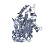
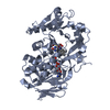
- Assembly
| Deposited unit | 
| ||||||||
|---|---|---|---|---|---|---|---|---|---|
| 1 | 
| ||||||||
| 2 | 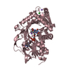
| ||||||||
| Unit cell |
|
-Components
-Protein , 1 types, 2 molecules AC
| #1: Protein | Mass: 49057.516 Da / Num. of mol.: 2 / Fragment: UNP residues 1-431 / Mutation: N56A Source method: isolated from a genetically manipulated source Source: (gene. exp.) Phaeosphaeria nodorum (strain SN15 / ATCC MYA-4574 / FGSC 10173) (fungus)Strain: SN15 / ATCC MYA-4574 / FGSC 10173 / Gene: SNOG_08398 / Plasmid: pET22b / Production host:  |
|---|
-Non-polymers , 5 types, 776 molecules 


| #2: Chemical |  Mass: 785.550 Da / Num. of mol.: 2 / Source method: obtained synthetically / Formula: C27H33N9O15P2 / Comment: FAD*YM Mass: 785.550 Da / Num. of mol.: 2 / Source method: obtained synthetically / Formula: C27H33N9O15P2 / Comment: FAD*YM#3: Chemical |  Mass: 60.052 Da / Num. of mol.: 2 / Source method: obtained synthetically / Formula: C2H4O2 Mass: 60.052 Da / Num. of mol.: 2 / Source method: obtained synthetically / Formula: C2H4O2#4: Chemical | ChemComp-CL /  Mass: 35.453 Da / Num. of mol.: 5 / Source method: obtained synthetically / Formula: Cl Mass: 35.453 Da / Num. of mol.: 5 / Source method: obtained synthetically / Formula: Cl#5: Chemical | ChemComp-NA /  Mass: 22.990 Da / Num. of mol.: 4 / Source method: obtained synthetically / Formula: Na Mass: 22.990 Da / Num. of mol.: 4 / Source method: obtained synthetically / Formula: Na#6: Water | ChemComp-HOH / | Mass: 18.015 Da / Num. of mol.: 763 / Source method: isolated from a natural source / Formula: H2O |
|---|
-Details
| Has protein modification | Y |
|---|
-Experimental details
-Experiment
| Experiment | Method: X-RAY DIFFRACTION / Number of used crystals: 1 |
|---|
- Sample preparation
| Crystal | Density Matthews: 2.44 Å3/Da / Density % sol: 49.63 % |
|---|---|
| Crystal grow | Temperature: 293 K / Method: vapor diffusion, sitting drop / pH: 5 / Details: Tacsimate, PEG 3350 |
-Data collection
| Diffraction | Mean temperature: 100 K | |||||||||||||||||||||||||||||||||||||||||||||||||||||||||||||||||||||||||||||
|---|---|---|---|---|---|---|---|---|---|---|---|---|---|---|---|---|---|---|---|---|---|---|---|---|---|---|---|---|---|---|---|---|---|---|---|---|---|---|---|---|---|---|---|---|---|---|---|---|---|---|---|---|---|---|---|---|---|---|---|---|---|---|---|---|---|---|---|---|---|---|---|---|---|---|---|---|---|---|
| Diffraction source | Source: ROTATING ANODE / Type: RIGAKU MICROMAX-007 HF / Wavelength: 1.5418 Å | |||||||||||||||||||||||||||||||||||||||||||||||||||||||||||||||||||||||||||||
| Detector | Type: RIGAKU RAXIS VII / Detector: IMAGE PLATE / Date: Dec 13, 2016 | |||||||||||||||||||||||||||||||||||||||||||||||||||||||||||||||||||||||||||||
| Radiation | Protocol: SINGLE WAVELENGTH / Monochromatic (M) / Laue (L): M / Scattering type: x-ray | |||||||||||||||||||||||||||||||||||||||||||||||||||||||||||||||||||||||||||||
| Radiation wavelength | Wavelength: 1.5418 Å / Relative weight: 1 | |||||||||||||||||||||||||||||||||||||||||||||||||||||||||||||||||||||||||||||
| Reflection | Resolution: 1.8→19.64 Å / Num. obs: 80576 / % possible obs: 96 % / Redundancy: 3.58 % / Rmerge(I) obs: 0.08 / Rrim(I) all: 0.08 / Χ2: 0.97 / Net I/σ(I): 6.6 / Num. measured all: 290996 / Scaling rejects: 2183 | |||||||||||||||||||||||||||||||||||||||||||||||||||||||||||||||||||||||||||||
| Reflection shell |
|
- Processing
| Software |
| ||||||||||||||||||||||||||||||||||||||||||||||||||||||||||||
|---|---|---|---|---|---|---|---|---|---|---|---|---|---|---|---|---|---|---|---|---|---|---|---|---|---|---|---|---|---|---|---|---|---|---|---|---|---|---|---|---|---|---|---|---|---|---|---|---|---|---|---|---|---|---|---|---|---|---|---|---|---|
| Refinement | Method to determine structure: MOLECULAR REPLACEMENT Starting model: 5T1F Resolution: 1.8→19.64 Å / Cor.coef. Fo:Fc: 0.963 / Cor.coef. Fo:Fc free: 0.943 / SU B: 4.062 / SU ML: 0.12 / Cross valid method: THROUGHOUT / σ(F): 0 / ESU R: 0.157 / ESU R Free: 0.155 Details: HYDROGENS HAVE BEEN ADDED IN THE RIDING POSITIONS U VALUES : REFINED INDIVIDUALLY
| ||||||||||||||||||||||||||||||||||||||||||||||||||||||||||||
| Solvent computation | Ion probe radii: 0.8 Å / Shrinkage radii: 0.8 Å / VDW probe radii: 1.2 Å | ||||||||||||||||||||||||||||||||||||||||||||||||||||||||||||
| Displacement parameters | Biso max: 136.36 Å2 / Biso mean: 32.891 Å2 / Biso min: 15.33 Å2
| ||||||||||||||||||||||||||||||||||||||||||||||||||||||||||||
| Refinement step | Cycle: final / Resolution: 1.8→19.64 Å
| ||||||||||||||||||||||||||||||||||||||||||||||||||||||||||||
| Refine LS restraints |
| ||||||||||||||||||||||||||||||||||||||||||||||||||||||||||||
| LS refinement shell | Resolution: 1.8→1.846 Å / Rfactor Rfree error: 0 / Total num. of bins used: 20
|
