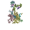
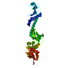
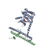
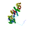
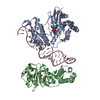
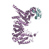
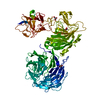
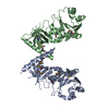
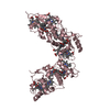
Journal: Nat Microbiol / Year: 2017 Title: Structural basis for λN-dependent processive transcription antitermination. Authors: Nelly Said / Ferdinand Krupp / Ekaterina Anedchenko / Karine F Santos / Olexandr Dybkov / Yong-Heng Huang / Chung-Tien Lee / Bernhard Loll / Elmar Behrmann / Jörg Bürger / Thorsten Mielke ...Authors: Nelly Said / Ferdinand Krupp / Ekaterina Anedchenko / Karine F Santos / Olexandr Dybkov / Yong-Heng Huang / Chung-Tien Lee / Bernhard Loll / Elmar Behrmann / Jörg Bürger / Thorsten Mielke / Justus Loerke / Henning Urlaub / Christian M T Spahn / Gert Weber / Markus C Wahl / Abstract: λN-mediated processive antitermination constitutes a paradigmatic transcription regulatory event, during which phage protein λN, host factors NusA, NusB, NusE and NusG, and an RNA nut site render ...λN-mediated processive antitermination constitutes a paradigmatic transcription regulatory event, during which phage protein λN, host factors NusA, NusB, NusE and NusG, and an RNA nut site render elongating RNA polymerase termination-resistant. The structural basis of the process has so far remained elusive. Here we describe a crystal structure of a λN-NusA-NusB-NusE-nut site complex and an electron cryo-microscopic structure of a complete transcription antitermination complex, comprising RNA polymerase, DNA, nut site RNA, all Nus factors and λN, validated by crosslinking/mass spectrometry. Due to intrinsic disorder, λN can act as a multiprotein/RNA interaction hub, which, together with nut site RNA, arranges NusA, NusB and NusE into a triangular complex. This complex docks via the NusA N-terminal domain and the λN C-terminus next to the RNA exit channel on RNA polymerase. Based on the structures, comparative crosslinking analyses and structure-guided mutagenesis, we hypothesize that λN mounts a multipronged strategy to reprogram the transcriptional machinery, which may include (1) the λN C terminus clamping the RNA exit channel, thus stabilizing the DNA:RNA hybrid; (2) repositioning of NusA and RNAP elements, thus redirecting nascent RNA and sequestering the upstream branch of a terminator hairpin; and (3) hindering RNA engagement of termination factor ρ and/or obstructing ρ translocation on the transcript.
In the structure databanks used in Yorodumi, some data are registered as the other names, "COVID-19 virus" and "2019-nCoV". Here are the details of the virus and the list of structure data.
Jan 31, 2019. EMDB accession codes are about to change! (news from PDBe EMDB page)
EMDB accession codes are about to change! (news from PDBe EMDB page)
The allocation of 4 digits for EMDB accession codes will soon come to an end. Whilst these codes will remain in use, new EMDB accession codes will include an additional digit and will expand incrementally as the available range of codes is exhausted. The current 4-digit format prefixed with “EMD-” (i.e. EMD-XXXX) will advance to a 5-digit format (i.e. EMD-XXXXX), and so on. It is currently estimated that the 4-digit codes will be depleted around Spring 2019, at which point the 5-digit format will come into force.
The EM Navigator/Yorodumi systems omit the EMD- prefix.
Related info.:Q: What is EMD? / ID/Accession-code notation in Yorodumi/EM Navigator
Yorodumi is a browser for structure data from EMDB, PDB, SASBDB, etc.
This page is also the successor to EM Navigator detail page, and also detail information page/front-end page for Omokage search.
The word "yorodu" (or yorozu) is an old Japanese word meaning "ten thousand". "mi" (miru) is to see.
Related info.:EMDB / PDB / SASBDB / Comparison of 3 databanks / Yorodumi Search / Aug 31, 2016. New EM Navigator & Yorodumi / Yorodumi Papers / Jmol/JSmol / Function and homology information / Changes in new EM Navigator and Yorodumi
 Movie
Movie Controller


 Open data
 Basic information
 Components
 Keywords
 Function and homology information
 X-RAY DIFFRACTION /
  Authors
 Citation
 Structure visualization
 Downloads & links
 Other downloads
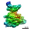


 PDBj
PDBj


 Assembly
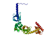

 Mass: 96.063 Da / Num. of mol.: 5 / Source method: obtained synthetically / Formula: SO4
 Mass: 24.305 Da / Num. of mol.: 1 / Source method: obtained synthetically / Formula: Mg
 Mass: 18.015 Da / Num. of mol.: 97 / Source method: isolated from a natural source / Formula: H2O
 Sample preparation
 Processing