 Movie
Movie Controller

[English] 日本語
 Yorodumi
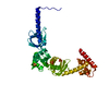
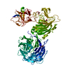
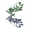
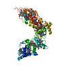
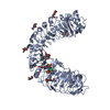
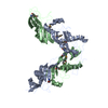
- PDB-6ao5: Crystal structure of human MST2 in complex with SAV1 SARAH domain -
+ Open data

- Basic information
| Entry | Database: PDB / ID: 6ao5 | ||||||
|---|---|---|---|---|---|---|---|
| Title | Crystal structure of human MST2 in complex with SAV1 SARAH domain | ||||||
 Components Components |
| ||||||
 Keywords Keywords | SIGNALING PROTEIN / Hippo / mst autoactivation / dimerization | ||||||
| Function / homology |  Function and homology information Function and homology informationkeratinocyte apoptotic process / regulation of cell differentiation involved in embryonic placenta development / primitive hemopoiesis / cell differentiation involved in embryonic placenta development / negative regulation of cell growth involved in contact inhibition / intestinal epithelial cell differentiation / neural tube formation / positive regulation of extrinsic apoptotic signaling pathway via death domain receptors / positive regulation of keratinocyte apoptotic process / regulation of stem cell population maintenance ...keratinocyte apoptotic process / regulation of cell differentiation involved in embryonic placenta development / primitive hemopoiesis / cell differentiation involved in embryonic placenta development / negative regulation of cell growth involved in contact inhibition / intestinal epithelial cell differentiation / neural tube formation / positive regulation of extrinsic apoptotic signaling pathway via death domain receptors / positive regulation of keratinocyte apoptotic process / regulation of stem cell population maintenance / positive regulation of hippo signaling / negative regulation of cardiac muscle cell proliferation / endocardium development / lung epithelial cell differentiation / negative regulation of organ growth / hippo signaling / transcription regulator activator activity / Signaling by Hippo / protein localization to centrosome / organ growth / ventricular septum morphogenesis / hepatocyte apoptotic process / regulation of MAPK cascade / extrinsic apoptotic signaling pathway via death domain receptors / hair follicle development / cardiac muscle cell proliferation / canonical Wnt signaling pathway / positive regulation of fat cell differentiation / keratinocyte differentiation / JNK cascade / epithelial cell proliferation / protein serine/threonine kinase activator activity / central nervous system development / protein tetramerization / phosphatidylinositol 3-kinase/protein kinase B signal transduction / negative regulation of canonical Wnt signaling pathway / positive regulation of JNK cascade / negative regulation of epithelial cell proliferation / protein import into nucleus / molecular adaptor activity / protein phosphorylation / protein kinase activity / non-specific serine/threonine protein kinase / positive regulation of phosphatidylinositol 3-kinase/protein kinase B signal transduction / intracellular signal transduction / protein stabilization / positive regulation of apoptotic process / negative regulation of cell population proliferation / protein serine kinase activity / protein serine/threonine kinase activity / apoptotic process / centrosome / magnesium ion binding / protein-containing complex / ATP binding / identical protein binding / nucleus / cytoplasm / cytosol Similarity search - Function | ||||||
| Biological species |  Homo sapiens (human) Homo sapiens (human) | ||||||
| Method |  X-RAY DIFFRACTION / SYNCHROTRON / MOLECULAR REPLACEMENT / Resolution: 2.955 Å X-RAY DIFFRACTION / SYNCHROTRON / MOLECULAR REPLACEMENT / Resolution: 2.955 Å | ||||||
 Authors Authors | Tomchick, D.R. / Luo, X. / Ni, L. | ||||||
 Citation Citation | Journal: Elife / Year: 2017 Title: SAV1 promotes Hippo kinase activation through antagonizing the PP2A phosphatase STRIPAK. Authors: Bae, S.J. / Ni, L. / Osinski, A. / Tomchick, D.R. / Brautigam, C.A. / Luo, X. #1: Journal: Structure / Year: 2013Title: Structural basis for autoactivation of human Mst2 kinase and its regulation by RASSF5. Authors: Ni, L. / Li, S. / Yu, J. / Min, J. / Brautigam, C.A. / Tomchick, D.R. / Pan, D. / Luo, X. | ||||||
| History |
|
- Structure visualization
| Structure viewer | Molecule: MolmilJmol/JSmol |
|---|
- Downloads & links
-Download
| PDBx/mmCIF format | 6ao5.cif.gz | 256.5 KB | Display | PDBx/mmCIF format |
|---|---|---|---|---|
| PDB format | pdb6ao5.ent.gz | 211.4 KB | Display | PDB format |
| PDBx/mmJSON format | 6ao5.json.gz | Tree view | PDBx/mmJSON format | |
| Others |  Other downloads Other downloads |
-Validation report
| Arichive directory | https://data.pdbj.org/pub/pdb/validation_reports/ao/6ao5ftp://data.pdbj.org/pub/pdb/validation_reports/ao/6ao5 | HTTPS FTP |
|---|
-Related structure data
| Related structure data | 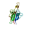 6ar0C  6ar2C  4lgdS S: Starting model for refinement C: citing same article ( |
|---|---|
| Similar structure data |
-Links
 PDBj
PDBj


- Assembly
| Deposited unit | 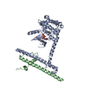
| ||||||||
|---|---|---|---|---|---|---|---|---|---|
| 1 |
| ||||||||
| Unit cell |
|
-Components
| #1: Protein | Mass: 41733.961 Da / Num. of mol.: 1 / Fragment: kinase domain (UNP residues 16-313, 428-491) / Mutation: D146N Source method: isolated from a genetically manipulated source Source: (gene. exp.) Homo sapiens (human) / Gene: STK3, KRS1, MST2 / Plasmid: PET29 / Production host:  References: UniProt: Q13188, non-specific serine/threonine protein kinase |
|---|---|
| #2: Protein | Mass: 11270.966 Da / Num. of mol.: 1 / Fragment: SARAH domain (UNP residues 291-383) Source method: isolated from a genetically manipulated source Source: (gene. exp.) Homo sapiens (human) / Gene: SAV1, WW45 / Plasmid: PET29 / Production host: |
| #3: Chemical | ChemComp-ANP /   Mass: 506.196 Da / Num. of mol.: 1 / Source method: obtained synthetically / Formula: C10H17N6O12P3 / Comment: AMP-PNP, energy-carrying molecule analogue*YM Mass: 506.196 Da / Num. of mol.: 1 / Source method: obtained synthetically / Formula: C10H17N6O12P3 / Comment: AMP-PNP, energy-carrying molecule analogue*YM |
| #4: Chemical | ChemComp-MG /   Mass: 24.305 Da / Num. of mol.: 1 / Source method: obtained synthetically / Formula: Mg Mass: 24.305 Da / Num. of mol.: 1 / Source method: obtained synthetically / Formula: Mg |
-Experimental details
-Experiment
| Experiment | Method: X-RAY DIFFRACTION / Number of used crystals: 1 |
|---|
- Sample preparation
| Crystal | Density Matthews: 3.62 Å3/Da / Density % sol: 65.99 % / Mosaicity: 0.579 ° |
|---|---|
| Crystal grow | Temperature: 293 K / Method: vapor diffusion, hanging drop Details: 0.05 M NaCl, 0.1 M Hepes, 0.19 mM CYMAL-7, 1 mM TCEP, 40% PEG 400 |
-Data collection
| Diffraction | Mean temperature: 100 K | |||||||||||||||||||||||||||||||||||||||||||||||||||||||||||||||||||||||||||||||||||||||||||||||||||||||||||||||||||||||||||||||||||||||||||||||||||||||||||||||||||||||||||||||||||||||||||||
|---|---|---|---|---|---|---|---|---|---|---|---|---|---|---|---|---|---|---|---|---|---|---|---|---|---|---|---|---|---|---|---|---|---|---|---|---|---|---|---|---|---|---|---|---|---|---|---|---|---|---|---|---|---|---|---|---|---|---|---|---|---|---|---|---|---|---|---|---|---|---|---|---|---|---|---|---|---|---|---|---|---|---|---|---|---|---|---|---|---|---|---|---|---|---|---|---|---|---|---|---|---|---|---|---|---|---|---|---|---|---|---|---|---|---|---|---|---|---|---|---|---|---|---|---|---|---|---|---|---|---|---|---|---|---|---|---|---|---|---|---|---|---|---|---|---|---|---|---|---|---|---|---|---|---|---|---|---|---|---|---|---|---|---|---|---|---|---|---|---|---|---|---|---|---|---|---|---|---|---|---|---|---|---|---|---|---|---|---|---|---|
| Diffraction source | Source: SYNCHROTRON / Site: APS  / Beamline: 19-ID / Wavelength: 0.97918 Å / Beamline: 19-ID / Wavelength: 0.97918 Å | |||||||||||||||||||||||||||||||||||||||||||||||||||||||||||||||||||||||||||||||||||||||||||||||||||||||||||||||||||||||||||||||||||||||||||||||||||||||||||||||||||||||||||||||||||||||||||||
| Detector | Type: ADSC QUANTUM 315r / Detector: CCD / Date: Oct 12, 2015 / Details: monochromator | |||||||||||||||||||||||||||||||||||||||||||||||||||||||||||||||||||||||||||||||||||||||||||||||||||||||||||||||||||||||||||||||||||||||||||||||||||||||||||||||||||||||||||||||||||||||||||||
| Radiation | Protocol: SINGLE WAVELENGTH / Monochromatic (M) / Laue (L): M / Scattering type: x-ray | |||||||||||||||||||||||||||||||||||||||||||||||||||||||||||||||||||||||||||||||||||||||||||||||||||||||||||||||||||||||||||||||||||||||||||||||||||||||||||||||||||||||||||||||||||||||||||||
| Radiation wavelength | Wavelength: 0.97918 Å / Relative weight: 1 | |||||||||||||||||||||||||||||||||||||||||||||||||||||||||||||||||||||||||||||||||||||||||||||||||||||||||||||||||||||||||||||||||||||||||||||||||||||||||||||||||||||||||||||||||||||||||||||
| Reflection | Resolution: 2.95→50 Å / Num. obs: 15976 / % possible obs: 99.9 % / Redundancy: 19 % / Biso Wilson estimate: 48.45 Å2 / Rmerge(I) obs: 0.124 / Rpim(I) all: 0.029 / Rrim(I) all: 0.128 / Χ2: 1.085 / Net I/σ(I): 7 / Num. measured all: 303392 | |||||||||||||||||||||||||||||||||||||||||||||||||||||||||||||||||||||||||||||||||||||||||||||||||||||||||||||||||||||||||||||||||||||||||||||||||||||||||||||||||||||||||||||||||||||||||||||
| Reflection shell | Diffraction-ID: 1
|
- Processing
| Software |
| |||||||||||||||||||||||||||||||||||||||||||||||||||||||||||||||||||||||||||||||||||||||||||||||||||||||||||||||||||||||||||||||||||||||||||||||||||||||||||||||||||||||||||||||
|---|---|---|---|---|---|---|---|---|---|---|---|---|---|---|---|---|---|---|---|---|---|---|---|---|---|---|---|---|---|---|---|---|---|---|---|---|---|---|---|---|---|---|---|---|---|---|---|---|---|---|---|---|---|---|---|---|---|---|---|---|---|---|---|---|---|---|---|---|---|---|---|---|---|---|---|---|---|---|---|---|---|---|---|---|---|---|---|---|---|---|---|---|---|---|---|---|---|---|---|---|---|---|---|---|---|---|---|---|---|---|---|---|---|---|---|---|---|---|---|---|---|---|---|---|---|---|---|---|---|---|---|---|---|---|---|---|---|---|---|---|---|---|---|---|---|---|---|---|---|---|---|---|---|---|---|---|---|---|---|---|---|---|---|---|---|---|---|---|---|---|---|---|---|---|---|---|
| Refinement | Method to determine structure: MOLECULAR REPLACEMENT Starting model: 4LGD Resolution: 2.955→42.271 Å / SU ML: 0.32 / Cross valid method: FREE R-VALUE / σ(F): 1.36 / Phase error: 28.54 / Stereochemistry target values: ML
| |||||||||||||||||||||||||||||||||||||||||||||||||||||||||||||||||||||||||||||||||||||||||||||||||||||||||||||||||||||||||||||||||||||||||||||||||||||||||||||||||||||||||||||||
| Solvent computation | Shrinkage radii: 0.9 Å / VDW probe radii: 1.11 Å / Solvent model: FLAT BULK SOLVENT MODEL | |||||||||||||||||||||||||||||||||||||||||||||||||||||||||||||||||||||||||||||||||||||||||||||||||||||||||||||||||||||||||||||||||||||||||||||||||||||||||||||||||||||||||||||||
| Displacement parameters | Biso max: 192.97 Å2 / Biso mean: 60.0404 Å2 / Biso min: 15.87 Å2 | |||||||||||||||||||||||||||||||||||||||||||||||||||||||||||||||||||||||||||||||||||||||||||||||||||||||||||||||||||||||||||||||||||||||||||||||||||||||||||||||||||||||||||||||
| Refinement step | Cycle: final / Resolution: 2.955→42.271 Å
| |||||||||||||||||||||||||||||||||||||||||||||||||||||||||||||||||||||||||||||||||||||||||||||||||||||||||||||||||||||||||||||||||||||||||||||||||||||||||||||||||||||||||||||||
| Refine LS restraints |
| |||||||||||||||||||||||||||||||||||||||||||||||||||||||||||||||||||||||||||||||||||||||||||||||||||||||||||||||||||||||||||||||||||||||||||||||||||||||||||||||||||||||||||||||
| LS refinement shell | Refine-ID: X-RAY DIFFRACTION / Rfactor Rfree error: 0 / Total num. of bins used: 5
| |||||||||||||||||||||||||||||||||||||||||||||||||||||||||||||||||||||||||||||||||||||||||||||||||||||||||||||||||||||||||||||||||||||||||||||||||||||||||||||||||||||||||||||||
| Refinement TLS params. | Method: refined / Refine-ID: X-RAY DIFFRACTION
| |||||||||||||||||||||||||||||||||||||||||||||||||||||||||||||||||||||||||||||||||||||||||||||||||||||||||||||||||||||||||||||||||||||||||||||||||||||||||||||||||||||||||||||||
| Refinement TLS group |
|
