 Movie
Movie Controller


+ Open data

- Basic information
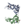
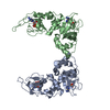
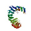
| Entry | Database: PDB / ID: 4z63 | ||||||
|---|---|---|---|---|---|---|---|
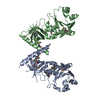
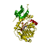
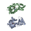
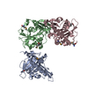
| Title | The plant peptide hormone receptor in arabidopsis | ||||||
 Components Components |
| ||||||
 Keywords Keywords | HORMONE / hormone receptor | ||||||
| Function / homology |  Function and homology information Function and homology informationregulation of defense response / peptide receptor activity / guanylate cyclase / guanylate cyclase activity / response to bacterium / protein kinase activity / non-specific serine/threonine protein kinase / innate immune response / protein serine kinase activity / protein serine/threonine kinase activity ...regulation of defense response / peptide receptor activity / guanylate cyclase / guanylate cyclase activity / response to bacterium / protein kinase activity / non-specific serine/threonine protein kinase / innate immune response / protein serine kinase activity / protein serine/threonine kinase activity / ATP binding / plasma membrane Similarity search - Function | ||||||
| Biological species |  | ||||||
| Method |  X-RAY DIFFRACTION / SYNCHROTRON / MOLECULAR REPLACEMENT / Resolution: 2.514 Å X-RAY DIFFRACTION / SYNCHROTRON / MOLECULAR REPLACEMENT / Resolution: 2.514 Å | ||||||
 Authors Authors | Chai, J. / Wang, J. / Han, Z. | ||||||
 Citation Citation | Journal: Nature / Year: 2015 Title: Allosteric receptor activation by the plant peptide hormone phytosulfokine Authors: Wang, J. / Li, H. / Han, Z. / Zhang, H. / Wang, T. / Lin, G. / Chang, J. / Yang, W. / Chai, J. | ||||||
| History |
|
- Structure visualization
| Structure viewer | Molecule: MolmilJmol/JSmol |
|---|
- Downloads & links
-Download
| PDBx/mmCIF format | 4z63.cif.gz | 140.5 KB | Display | PDBx/mmCIF format |
|---|---|---|---|---|
| PDB format | pdb4z63.ent.gz | 107.1 KB | Display | PDB format |
| PDBx/mmJSON format | 4z63.json.gz | Tree view | PDBx/mmJSON format | |
| Others |  Other downloads Other downloads |
-Validation report
| Arichive directory | https://data.pdbj.org/pub/pdb/validation_reports/z6/4z63ftp://data.pdbj.org/pub/pdb/validation_reports/z6/4z63 | HTTPS FTP |
|---|
-Related structure data
| Related structure data |  4z5wSC  4z61C  4z62C 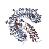 4z64C S: Starting model for refinement C: citing same article ( |
|---|---|
| Similar structure data |

-Links
 PDBj
PDBj


- Assembly
| Deposited unit | 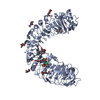
| ||||||||
|---|---|---|---|---|---|---|---|---|---|
| 1 |
| ||||||||
| Unit cell |
|
-Components
| #1: Protein | Mass: 69884.008 Da / Num. of mol.: 1 / Fragment: UNP residues 24-648 Source method: isolated from a genetically manipulated source Source: (gene. exp.) Production host: Insect cell expression vector pTIE1 (others) References: UniProt: Q9ZVR7, non-specific serine/threonine protein kinase | ||||
|---|---|---|---|---|---|
| #2: Protein/peptide | Mass: 846.879 Da / Num. of mol.: 1 / Source method: obtained synthetically / Source: (synth.) | ||||
| #3: Sugar | ChemComp-NAG /   Type: D-saccharide, beta linking / Mass: 221.208 Da / Num. of mol.: 9 Type: D-saccharide, beta linking / Mass: 221.208 Da / Num. of mol.: 9Source method: isolated from a genetically manipulated source Formula: C8H15NO6 #4: Water | ChemComp-HOH / |  Mass: 18.015 Da / Num. of mol.: 145 / Source method: isolated from a natural source / Formula: H2O Mass: 18.015 Da / Num. of mol.: 145 / Source method: isolated from a natural source / Formula: H2OHas protein modification | Y | |
-Experimental details
-Experiment
| Experiment | Method: X-RAY DIFFRACTION / Number of used crystals: 1 |
|---|
- Sample preparation
| Crystal | Density Matthews: 3.84 Å3/Da / Density % sol: 67.93 % |
|---|---|
| Crystal grow | Temperature: 291 K / Method: vapor diffusion, hanging drop / Details: 0.1M Bis-Tris 5.5, 2.0M (NH4)2SO4 |
-Data collection
| Diffraction | Mean temperature: 100 K |
|---|---|
| Diffraction source | Source: SYNCHROTRON / Site: SSRF  / Beamline: BL17U / Wavelength: 1 Å / Beamline: BL17U / Wavelength: 1 Å |
| Detector | Type: BRUKER SMART 6500 / Detector: CCD / Date: Dec 9, 2013 |
| Radiation | Protocol: SINGLE WAVELENGTH / Monochromatic (M) / Laue (L): M / Scattering type: x-ray |
| Radiation wavelength | Wavelength: 1 Å / Relative weight: 1 |
| Reflection | Resolution: 2.5→99 Å / Num. obs: 36369 / % possible obs: 98.4 % / Redundancy: 5.3 % / Net I/σ(I): 26 |
- Processing
| Software |
| ||||||||||||||||||||||||||||||||||||||||||||||||||||||||||||||||||||||||||||||||||||||||||||||||||
|---|---|---|---|---|---|---|---|---|---|---|---|---|---|---|---|---|---|---|---|---|---|---|---|---|---|---|---|---|---|---|---|---|---|---|---|---|---|---|---|---|---|---|---|---|---|---|---|---|---|---|---|---|---|---|---|---|---|---|---|---|---|---|---|---|---|---|---|---|---|---|---|---|---|---|---|---|---|---|---|---|---|---|---|---|---|---|---|---|---|---|---|---|---|---|---|---|---|---|---|
| Refinement | Method to determine structure: MOLECULAR REPLACEMENT Starting model: 4Z5W Resolution: 2.514→33.544 Å / SU ML: 0.34 / Cross valid method: THROUGHOUT / σ(F): 0 / Phase error: 29.28 / Stereochemistry target values: ML
| ||||||||||||||||||||||||||||||||||||||||||||||||||||||||||||||||||||||||||||||||||||||||||||||||||
| Solvent computation | Shrinkage radii: 0.9 Å / VDW probe radii: 1.11 Å / Solvent model: FLAT BULK SOLVENT MODEL | ||||||||||||||||||||||||||||||||||||||||||||||||||||||||||||||||||||||||||||||||||||||||||||||||||
| Refinement step | Cycle: LAST / Resolution: 2.514→33.544 Å
| ||||||||||||||||||||||||||||||||||||||||||||||||||||||||||||||||||||||||||||||||||||||||||||||||||
| Refine LS restraints |
| ||||||||||||||||||||||||||||||||||||||||||||||||||||||||||||||||||||||||||||||||||||||||||||||||||
| LS refinement shell |
|
