 Movie
Movie Controller

[English] 日本語
 Yorodumi
- PDB-7kua: Crystal structure of the MarR family transcriptional regulator fr... -
+ Open data

- Basic information
| Entry | Database: PDB / ID: 7kua | ||||||
|---|---|---|---|---|---|---|---|
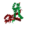
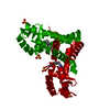
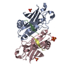
| Title | Crystal structure of the MarR family transcriptional regulator from Pseudomonas putida bound to Indole 3 acetic acid | ||||||
 Components Components | Transcriptional regulator, MarR family | ||||||
 Keywords Keywords | DNA BINDING PROTEIN / Transcriptional Regulator / Ligand Binding | ||||||
| Function / homology |  Function and homology information Function and homology informationresponse to stress / DNA-binding transcription factor activity / DNA binding Similarity search - Function | ||||||
| Biological species |  Pseudomonas putida (bacteria) Pseudomonas putida (bacteria) | ||||||
| Method |  X-RAY DIFFRACTION / SYNCHROTRON / MOLECULAR REPLACEMENT / Resolution: 1.89 Å X-RAY DIFFRACTION / SYNCHROTRON / MOLECULAR REPLACEMENT / Resolution: 1.89 Å | ||||||
 Authors Authors | Walton, W.G. / Lietzan, A.D. / Redinbo, M.R. / Dangl, J.L. | ||||||
| Funding support |  United States, 1items United States, 1items
| ||||||
 Citation Citation | Journal: Nat Microbiol / Year: 2022 Title: Diverse MarR bacterial regulators of auxin catabolism in the plant microbiome. Authors: Conway, J.M. / Walton, W.G. / Salas-Gonzalez, I. / Law, T.F. / Lindberg, C.A. / Crook, L.E. / Kosina, S.M. / Fitzpatrick, C.R. / Lietzan, A.D. / Northen, T.R. / Jones, C.D. / Finkel, O.M. / ...Authors: Conway, J.M. / Walton, W.G. / Salas-Gonzalez, I. / Law, T.F. / Lindberg, C.A. / Crook, L.E. / Kosina, S.M. / Fitzpatrick, C.R. / Lietzan, A.D. / Northen, T.R. / Jones, C.D. / Finkel, O.M. / Redinbo, M.R. / Dangl, J.L. | ||||||
| History |
|
- Structure visualization
| Structure viewer | Molecule: MolmilJmol/JSmol |
|---|
- Downloads & links
-Download
| PDBx/mmCIF format | 7kua.cif.gz | 53.4 KB | Display | PDBx/mmCIF format |
|---|---|---|---|---|
| PDB format | pdb7kua.ent.gz | 32.4 KB | Display | PDB format |
| PDBx/mmJSON format | 7kua.json.gz | Tree view | PDBx/mmJSON format | |
| Others |  Other downloads Other downloads |
-Validation report
| Arichive directory | https://data.pdbj.org/pub/pdb/validation_reports/ku/7kuaftp://data.pdbj.org/pub/pdb/validation_reports/ku/7kua | HTTPS FTP |
|---|
-Related structure data
| Related structure data |  7kfoC  7kfqC  7kfsC  7kh3C  7kigC  7kjlC  7kjqC  7kk0C  7kkcC  7kkiC  7krhC  7kymC  7l19C  7l1iC  3cjnS S: Starting model for refinement C: citing same article ( |
|---|---|
| Similar structure data |


-Links
 PDBj
PDBj
- Assembly
| Deposited unit | 
| ||||||||||||
|---|---|---|---|---|---|---|---|---|---|---|---|---|---|
| 1 | 
| ||||||||||||
| Unit cell |
|
-Components
| #1: Protein | Mass: 22120.930 Da / Num. of mol.: 1 Source method: isolated from a genetically manipulated source Source: (gene. exp.) Pseudomonas putida (bacteria) / Gene: iacR, E6B08_12615 / Production host: |
|---|---|
| #2: Chemical | ChemComp-IAC /   Mass: 175.184 Da / Num. of mol.: 1 / Source method: obtained synthetically / Formula: C10H9NO2 / Feature type: SUBJECT OF INVESTIGATION / Comment: hormone*YM Mass: 175.184 Da / Num. of mol.: 1 / Source method: obtained synthetically / Formula: C10H9NO2 / Feature type: SUBJECT OF INVESTIGATION / Comment: hormone*YM |
| #3: Water | ChemComp-HOH /  Mass: 18.015 Da / Num. of mol.: 58 / Source method: isolated from a natural source / Formula: H2O Mass: 18.015 Da / Num. of mol.: 58 / Source method: isolated from a natural source / Formula: H2O |
| Has ligand of interest | Y |
-Experimental details
-Experiment
| Experiment | Method: X-RAY DIFFRACTION / Number of used crystals: 1 |
|---|
- Sample preparation
| Crystal | Density Matthews: 1.98 Å3/Da / Density % sol: 37.73 % |
|---|---|
| Crystal grow | Temperature: 293 K / Method: vapor diffusion, sitting drop Details: Anatrace MCSG2 screen 35 % (v/v) Microlytic Mix(1), pH 7.0, which consists of: 1.8305 M Malonic acid, 0.25 M Ammonium citrate tribasic, 0.12 M Succinic acid, 0.3 M DL-Malic acid, 0.4 M ...Details: Anatrace MCSG2 screen 35 % (v/v) Microlytic Mix(1), pH 7.0, which consists of: 1.8305 M Malonic acid, 0.25 M Ammonium citrate tribasic, 0.12 M Succinic acid, 0.3 M DL-Malic acid, 0.4 M Sodium acetate trihydrate, 0.5 M Sodium formate, 0.16 M Ammonium tartrate dibasic, the mixture titrated to pH 7.0 using sodium hydroxide |
-Data collection
| Diffraction | Mean temperature: 100 K / Serial crystal experiment: N |
|---|---|
| Diffraction source | Source: SYNCHROTRON / Site: APS / Beamline: 23-ID-D / Wavelength: 1.03322 Å |
| Detector | Type: DECTRIS PILATUS3 6M / Detector: PIXEL / Date: Nov 15, 2020 |
| Radiation | Protocol: SINGLE WAVELENGTH / Monochromatic (M) / Laue (L): M / Scattering type: x-ray |
| Radiation wavelength | Wavelength: 1.03322 Å / Relative weight: 1 |
| Reflection | Resolution: 1.88→50 Å / Num. obs: 14591 / % possible obs: 99.9 % / Redundancy: 8.3 % / Biso Wilson estimate: 29.48 Å2 / CC1/2: 1 / Net I/σ(I): 33.4 |
| Reflection shell | Resolution: 1.88→1.91 Å / Num. unique obs: 715 / CC1/2: 0.957 |
- Processing
| Software |
| |||||||||||||||||||||||||||||||||||||||||||||||||||||||||||||||||||||||||||||
|---|---|---|---|---|---|---|---|---|---|---|---|---|---|---|---|---|---|---|---|---|---|---|---|---|---|---|---|---|---|---|---|---|---|---|---|---|---|---|---|---|---|---|---|---|---|---|---|---|---|---|---|---|---|---|---|---|---|---|---|---|---|---|---|---|---|---|---|---|---|---|---|---|---|---|---|---|---|---|
| Refinement | Method to determine structure: MOLECULAR REPLACEMENT Starting model: 3CJN Resolution: 1.89→25.99 Å / SU ML: 0.2014 / Cross valid method: FREE R-VALUE / σ(F): 1.38 / Phase error: 26.0325 Stereochemistry target values: GeoStd + Monomer Library + CDL v1.2
| |||||||||||||||||||||||||||||||||||||||||||||||||||||||||||||||||||||||||||||
| Solvent computation | Shrinkage radii: 0.9 Å / VDW probe radii: 1.11 Å / Solvent model: FLAT BULK SOLVENT MODEL | |||||||||||||||||||||||||||||||||||||||||||||||||||||||||||||||||||||||||||||
| Displacement parameters | Biso mean: 39.08 Å2 | |||||||||||||||||||||||||||||||||||||||||||||||||||||||||||||||||||||||||||||
| Refinement step | Cycle: LAST / Resolution: 1.89→25.99 Å
| |||||||||||||||||||||||||||||||||||||||||||||||||||||||||||||||||||||||||||||
| Refine LS restraints |
| |||||||||||||||||||||||||||||||||||||||||||||||||||||||||||||||||||||||||||||
| LS refinement shell |
|
