 Movie
Movie Controller

[English] 日本語
 Yorodumi
- PDB-1kbz: Crystal Structure of apo-dTDP-6-deoxy-L-lyxo-4-hexulose reductase... -
+ Open data

- Basic information
| Entry | Database: PDB / ID: 1kbz | ||||||
|---|---|---|---|---|---|---|---|
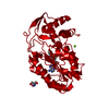
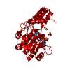
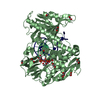
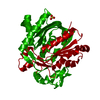
| Title | Crystal Structure of apo-dTDP-6-deoxy-L-lyxo-4-hexulose reductase (RmlD) from Salmonella enterica serovar Typhimurium | ||||||
 Components Components | dTDP-glucose oxidoreductase | ||||||
 Keywords Keywords | OXIDOREDUCTASE / Rossman-fold / sugar-nucleotide-binding domain | ||||||
| Function / homology |  Function and homology information Function and homology informationdTDP-4-dehydrorhamnose reductase / dTDP-4-dehydrorhamnose reductase activity / O antigen biosynthetic process / dTDP-rhamnose biosynthetic process / lipopolysaccharide biosynthetic process / polysaccharide biosynthetic process / metal ion binding / cytosol Similarity search - Function | ||||||
| Biological species |  Salmonella typhimurium (bacteria) Salmonella typhimurium (bacteria) | ||||||
| Method |  X-RAY DIFFRACTION / SYNCHROTRON / MAD / Resolution: 2.2 Å X-RAY DIFFRACTION / SYNCHROTRON / MAD / Resolution: 2.2 Å | ||||||
 Authors Authors | Blankenfeldt, W. / Kerr, I.D. / Giraud, M.F. / McMiken, H.J. / Leonard, G.A. / Whitfield, C. / Messner, P. / Graninger, M. / Naismith, J.H. | ||||||
 Citation Citation | Journal: Structure / Year: 2002 Title: Variation on a theme of SDR. dTDP-6-deoxy-L- lyxo-4-hexulose reductase (RmlD) shows a new Mg2+-dependent dimerization mode. Authors: Blankenfeldt, W. / Kerr, I.D. / Giraud, M.F. / McMiken, H.J. / Leonard, G. / Whitfield, C. / Messner, P. / Graninger, M. / Naismith, J.H. #1: Journal: Acta Crystallogr.,Sect.D / Year: 1999Title: Overexpression, purification, crystallisation and preliminary structural study of dTDP-6-deoxy-lyxo-4-reductase (RmlD) the fourth enzyme of the dTDP-rhanose synthesis pathway,from Salmonella ...Title: Overexpression, purification, crystallisation and preliminary structural study of dTDP-6-deoxy-lyxo-4-reductase (RmlD) the fourth enzyme of the dTDP-rhanose synthesis pathway,from Salmonella entrica serovar Typhimurim Authors: Giraud, M.F. / McMiken, H.J. / Leonard, G.A. / Messner, P. / Whitfield, C. / Naismith, J.H. #2: Journal: Curr.Opin.Struct.Biol. / Year: 2000Title: The rhamnose pathway Authors: Giraud, M.F. / Naismith, J.H. | ||||||
| History |
|
- Structure visualization
| Structure viewer | Molecule: MolmilJmol/JSmol |
|---|
- Downloads & links
-Download
| PDBx/mmCIF format | 1kbz.cif.gz | 74.5 KB | Display | PDBx/mmCIF format |
|---|---|---|---|---|
| PDB format | pdb1kbz.ent.gz | 55.3 KB | Display | PDB format |
| PDBx/mmJSON format | 1kbz.json.gz | Tree view | PDBx/mmJSON format | |
| Others |  Other downloads Other downloads |
-Validation report
| Arichive directory | https://data.pdbj.org/pub/pdb/validation_reports/kb/1kbzftp://data.pdbj.org/pub/pdb/validation_reports/kb/1kbz | HTTPS FTP |
|---|
-Related structure data


-Links
 PDBj
PDBj- Assembly
| Deposited unit | 
| ||||||||||||||||||
|---|---|---|---|---|---|---|---|---|---|---|---|---|---|---|---|---|---|---|---|
| 1 | 
| ||||||||||||||||||
| Unit cell |
| ||||||||||||||||||
| Components on special symmetry positions |
| ||||||||||||||||||
| Details | The biological assembly is a dimer generated by the the two-fold crystallographic c-axis: -x, -y, z. |
-Components
| #1: Protein | Mass: 32589.943 Da / Num. of mol.: 1 Source method: isolated from a genetically manipulated source Source: (gene. exp.) Salmonella typhimurium (bacteria) / Gene: rfbA / Plasmid: pET30 / Production host: References: UniProt: P26392, dTDP-4-dehydrorhamnose reductase |
|---|---|
| #2: Chemical | ChemComp-MG /   Mass: 24.305 Da / Num. of mol.: 1 / Source method: obtained synthetically / Formula: Mg Mass: 24.305 Da / Num. of mol.: 1 / Source method: obtained synthetically / Formula: Mg |
| #3: Water | ChemComp-HOH /  Mass: 18.015 Da / Num. of mol.: 171 / Source method: isolated from a natural source / Formula: H2O Mass: 18.015 Da / Num. of mol.: 171 / Source method: isolated from a natural source / Formula: H2O |
-Experimental details
-Experiment
| Experiment | Method: X-RAY DIFFRACTION / Number of used crystals: 1 |
|---|
- Sample preparation
| Crystal | Density Matthews: 2.15 Å3/Da / Density % sol: 42.87 % | ||||||||||||||||||||||||||||||||||||||||||
|---|---|---|---|---|---|---|---|---|---|---|---|---|---|---|---|---|---|---|---|---|---|---|---|---|---|---|---|---|---|---|---|---|---|---|---|---|---|---|---|---|---|---|---|
| Crystal grow | Temperature: 298 K / Method: vapor diffusion, sitting drop / pH: 8 Details: HEPES, PEG 4000, magnesium chloride, pH 8.0, VAPOR DIFFUSION, SITTING DROP at 298K | ||||||||||||||||||||||||||||||||||||||||||
| Crystal grow | *PLUS pH: 7.1 | ||||||||||||||||||||||||||||||||||||||||||
| Components of the solutions | *PLUS
|
-Data collection
| Diffraction | Mean temperature: 100 K |
|---|---|
| Diffraction source | Source: SYNCHROTRON / Site: ESRF  / Beamline: ID14-2 / Wavelength: 0.934 Å / Beamline: ID14-2 / Wavelength: 0.934 Å |
| Detector | Type: ADSC QUANTUM 4 / Detector: CCD / Date: May 1, 2001 |
| Radiation | Protocol: SINGLE WAVELENGTH / Monochromatic (M) / Laue (L): M / Scattering type: x-ray |
| Radiation wavelength | Wavelength: 0.934 Å / Relative weight: 1 |
| Reflection | Resolution: 2.21→54.23 Å / Num. all: 14547 / Num. obs: 14547 / % possible obs: 99.4 % / Observed criterion σ(I): 2.9 / Redundancy: 11.8 % / Biso Wilson estimate: 47 Å2 / Rmerge(I) obs: 0.072 / Net I/σ(I): 4.7 |
| Reflection shell | Resolution: 2.21→2.27 Å / Redundancy: 5.4 % / Rmerge(I) obs: 0.249 / Mean I/σ(I) obs: 2.9 / % possible all: 92 |
| Reflection | *PLUS Num. measured all: 171243 / Rmerge(I) obs: 0.072 |
| Reflection shell | *PLUS % possible obs: 92 % / Rmerge(I) obs: 0.249 |
- Processing
| Software |
| ||||||||||||||||||||||||||||
|---|---|---|---|---|---|---|---|---|---|---|---|---|---|---|---|---|---|---|---|---|---|---|---|---|---|---|---|---|---|
| Refinement | Method to determine structure: MAD / Resolution: 2.2→81.65 Å / SU B: 8.595 / SU ML: 0.224 / SU Rfree: 0.247 / Cross valid method: THROUGHOUT / σ(F): 0 / ESU R: 0.348 / ESU R Free: 0.247 / Details: HYDROGENS HAVE BEEN ADDED IN THE RIDING POSITIONS
| ||||||||||||||||||||||||||||
| Solvent computation | Solvent model: BABINET MODEL WITH MASK. PARAMETERS FOR MASK CALCULATION: VDW PROBE RADIUS: 1.40; ION PROBE RADIUS: 0.80; SHRINKAGE RADIUS: 0.80 | ||||||||||||||||||||||||||||
| Displacement parameters | Biso mean: 33.39 Å2
| ||||||||||||||||||||||||||||
| Refine analyze |
| ||||||||||||||||||||||||||||
| Refinement step | Cycle: LAST / Resolution: 2.2→81.65 Å
| ||||||||||||||||||||||||||||
| Refine LS restraints |
| ||||||||||||||||||||||||||||
| LS refinement shell | Resolution: 2.21→2.26 Å / Total num. of bins used: 20 /
| ||||||||||||||||||||||||||||
| Refinement | *PLUS Highest resolution: 2.21 Å / Rfactor all: 0.1939 / Rfactor obs: 0.192 / Rfactor Rfree: 0.256 / Rfactor Rwork: 0.189 | ||||||||||||||||||||||||||||
| Solvent computation | *PLUS | ||||||||||||||||||||||||||||
| Displacement parameters | *PLUS | ||||||||||||||||||||||||||||
| Refine LS restraints | *PLUS
| ||||||||||||||||||||||||||||
| LS refinement shell | *PLUS Rfactor Rfree: 0.229 / Rfactor Rwork: 0.223 |
