 Movie
Movie Controller

[English] 日本語
 Yorodumi
- PDB-7khf: CryoEM structure of LILRB1 D3D4 domain-inserted antibody MDB1 Fab... -
+ Open data

- Basic information
| Entry | Database: PDB / ID: 7khf | |||||||||
|---|---|---|---|---|---|---|---|---|---|---|
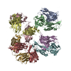
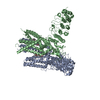
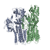
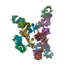
| Title | CryoEM structure of LILRB1 D3D4 domain-inserted antibody MDB1 Fab in complex with Plasmodium RIFIN (PF3D7_1373400) V2 domain | |||||||||
 Components Components |
| |||||||||
 Keywords Keywords | IMMUNE SYSTEM / LILRB1 / RIFIN / Malaria / Receptor-inserted antibody / MDB1 | |||||||||
| Function / homology | Variant surface antigen Rifin / Rifin / Immunoglobulins / Immunoglobulin-like / Sandwich / Mainly Beta / Rifin Function and homology information Function and homology information | |||||||||
| Biological species |  Homo sapiens (human) Homo sapiens (human) | |||||||||
| Method | ELECTRON MICROSCOPY / single particle reconstruction / cryo EM / Resolution: 3.54 Å | |||||||||
 Authors Authors | Xu, K. / Kwong, P.D. | |||||||||
| Funding support |  United States, 2items United States, 2items
| |||||||||
 Citation Citation | Journal: Nature / Year: 2021 Title: Structural basis of malaria RIFIN binding by LILRB1-containing antibodies. Authors: Yiwei Chen / Kai Xu / Luca Piccoli / Mathilde Foglierini / Joshua Tan / Wenjie Jin / Jason Gorman / Yaroslav Tsybovsky / Baoshan Zhang / Boubacar Traore / Chiara Silacci-Fregni / Claudia ...Authors: Yiwei Chen / Kai Xu / Luca Piccoli / Mathilde Foglierini / Joshua Tan / Wenjie Jin / Jason Gorman / Yaroslav Tsybovsky / Baoshan Zhang / Boubacar Traore / Chiara Silacci-Fregni / Claudia Daubenberger / Peter D Crompton / Roger Geiger / Federica Sallusto / Peter D Kwong / Antonio Lanzavecchia /  Abstract: Some Plasmodium falciparum repetitive interspersed families of polypeptides (RIFINs)-variant surface antigens that are expressed on infected erythrocytes-bind to the inhibitory receptor LAIR1, and ...Some Plasmodium falciparum repetitive interspersed families of polypeptides (RIFINs)-variant surface antigens that are expressed on infected erythrocytes-bind to the inhibitory receptor LAIR1, and insertion of DNA that encodes LAIR1 into immunoglobulin genes generates RIFIN-specific antibodies. Here we address the general relevance of this finding by searching for antibodies that incorporate LILRB1, another inhibitory receptor that binds to β2 microglobulin and RIFINs through their apical domains. By screening plasma from a cohort of donors from Mali, we identified individuals with LILRB1-containing antibodies. B cell clones isolated from three donors showed large DNA insertions in the switch region that encodes non-apical LILRB1 extracellular domain 3 and 4 (D3D4) or D3 alone in the variable-constant (VH-CH1) elbow. Through mass spectrometry and binding assays, we identified a large set of RIFINs that bind to LILRB1 D3. Crystal and cryo-electron microscopy structures of a RIFIN in complex with either LILRB1 D3D4 or a D3D4-containing antibody Fab revealed a mode of RIFIN-LILRB1 D3 interaction that is similar to that of RIFIN-LAIR1. The Fab showed an unconventional triangular architecture with the inserted LILRB1 domains opening up the VH-CH1 elbow without affecting VH-VL or CH1-CL pairing. Collectively, these findings show that RIFINs bind to LILRB1 through D3 and illustrate, with a naturally selected example, the general principle of creating novel antibodies by inserting receptor domains into the VH-CH1 elbow. | |||||||||
| History |
|
- Structure visualization
| Movie |
Movie viewer |
|---|---|
| Structure viewer | Molecule: MolmilJmol/JSmol |
- Downloads & links
-Download
| PDBx/mmCIF format | 7khf.cif.gz | 263.5 KB | Display | PDBx/mmCIF format |
|---|---|---|---|---|
| PDB format | pdb7khf.ent.gz | 212.9 KB | Display | PDB format |
| PDBx/mmJSON format | 7khf.json.gz | Tree view | PDBx/mmJSON format | |
| Others |  Other downloads Other downloads |
-Validation report
| Arichive directory | https://data.pdbj.org/pub/pdb/validation_reports/kh/7khfftp://data.pdbj.org/pub/pdb/validation_reports/kh/7khf | HTTPS FTP |
|---|
-Related structure data
| Related structure data |  22879MC  7kfkC C: citing same article ( M: map data used to model this data |
|---|---|
| Similar structure data |


-Links
 PDBj
PDBj

- Assembly
| Deposited unit | 
|
|---|---|
| 1 |
|
-Components
| #1: Antibody | Mass: 46384.973 Da / Num. of mol.: 2 Source method: isolated from a genetically manipulated source Source: (gene. exp.) Homo sapiens (human) / Production host: Homo sapiens (human)#2: Antibody | Mass: 22870.232 Da / Num. of mol.: 2 Source method: isolated from a genetically manipulated source Source: (gene. exp.) Homo sapiens (human) / Production host: Homo sapiens (human)#3: Protein | Mass: 15420.644 Da / Num. of mol.: 2 Source method: isolated from a genetically manipulated source Source: (gene. exp.) Strain: isolate 3D7 / Gene: PF3D7_1373400 / Production host: Homo sapiens (human) / References: UniProt: C0H5N9#4: Polysaccharide | 2-acetamido-2-deoxy-beta-D-glucopyranose-(1-4)-2-acetamido-2-deoxy-beta-D-glucopyranose Source method: isolated from a genetically manipulated source #5: Sugar |   Type: D-saccharide, beta linking / Mass: 221.208 Da / Num. of mol.: 2 / Source method: obtained synthetically / Formula: C8H15NO6 Type: D-saccharide, beta linking / Mass: 221.208 Da / Num. of mol.: 2 / Source method: obtained synthetically / Formula: C8H15NO6Has ligand of interest | N | Has protein modification | Y | |
|---|
-Experimental details
-Experiment
| Experiment | Method: ELECTRON MICROSCOPY |
|---|---|
| EM experiment | Aggregation state: PARTICLE / 3D reconstruction method: single particle reconstruction |
- Sample preparation
| Component |
| ||||||||||||||||||
|---|---|---|---|---|---|---|---|---|---|---|---|---|---|---|---|---|---|---|---|
| Molecular weight |
| ||||||||||||||||||
| Source (natural) |
| ||||||||||||||||||
| Source (recombinant) |
| ||||||||||||||||||
| Buffer solution | pH: 7.4 | ||||||||||||||||||
| Buffer component | Formula: PBS | ||||||||||||||||||
| Specimen | Conc.: 1 mg/ml / Embedding applied: NO / Shadowing applied: NO / Staining applied: NO / Vitrification applied: YES | ||||||||||||||||||
| Specimen support | Grid material: COPPER / Grid type: C-flat-1.2/1.3 | ||||||||||||||||||
| Vitrification | Instrument: FEI VITROBOT MARK IV / Cryogen name: ETHANE / Humidity: 90 % / Chamber temperature: 293 K |
- Electron microscopy imaging
| Experimental equipment |  Model: Titan Krios / Image courtesy: FEI Company |
|---|---|
| Microscopy | Model: FEI TITAN KRIOS |
| Electron gun | Electron source:  FIELD EMISSION GUN / Accelerating voltage: 300 kV / Illumination mode: FLOOD BEAM FIELD EMISSION GUN / Accelerating voltage: 300 kV / Illumination mode: FLOOD BEAM |
| Electron lens | Mode: BRIGHT FIELD / C2 aperture diameter: 70 µm |
| Specimen holder | Cryogen: NITROGEN / Specimen holder model: FEI TITAN KRIOS AUTOGRID HOLDER |
| Image recording | Average exposure time: 2 sec. / Electron dose: 70.72 e/Å2 / Film or detector model: GATAN K3 (6k x 4k) / Num. of grids imaged: 1 / Num. of real images: 6473 |
- Processing
| EM software |
| ||||||||||||||||||||||||||||||||||||
|---|---|---|---|---|---|---|---|---|---|---|---|---|---|---|---|---|---|---|---|---|---|---|---|---|---|---|---|---|---|---|---|---|---|---|---|---|---|
| CTF correction | Type: PHASE FLIPPING AND AMPLITUDE CORRECTION | ||||||||||||||||||||||||||||||||||||
| Particle selection | Num. of particles selected: 6010700 | ||||||||||||||||||||||||||||||||||||
| 3D reconstruction | Resolution: 3.54 Å / Resolution method: FSC 0.143 CUT-OFF / Num. of particles: 390908 / Symmetry type: POINT | ||||||||||||||||||||||||||||||||||||
| Atomic model building | Protocol: FLEXIBLE FIT / Space: REAL | ||||||||||||||||||||||||||||||||||||
| Atomic model building | PDB-ID: 4LLA Pdb chain-ID: A / Accession code: 4LLA / Source name: PDB / Type: experimental model |

