 Movie
Movie Controller

[English] 日本語
 Yorodumi
- EMDB-22879: cryoEM structure of LILRB1 D3D4 domain-inserted antibody MDB1 Fab... -
+ Open data

- Basic information
| Entry | Database: EMDB / ID: EMD-22879 | |||||||||
|---|---|---|---|---|---|---|---|---|---|---|
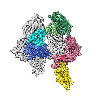
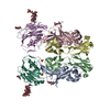
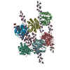
| Title | cryoEM structure of LILRB1 D3D4 domain-inserted antibody MDB1 Fab in complex with Plasmodium RIFIN (PF3D7_1373400) V2 domain | |||||||||
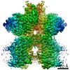
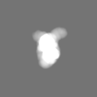
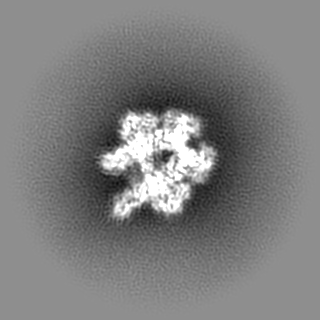
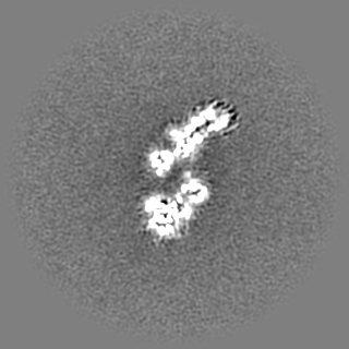
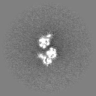
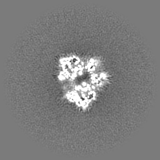
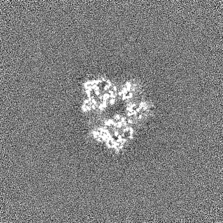
 Map data Map data | Primary sharpened map | |||||||||
 Sample Sample |
| |||||||||
 Keywords Keywords | LILRB1 / RIFIN / Malaria / Receptor-inserted antibody / MDB1 / IMMUNE SYSTEM | |||||||||
| Function / homology | Variant surface antigen Rifin / Rifin / Rifin Function and homology information Function and homology information | |||||||||
| Biological species |  Homo sapiens (human) / Homo sapiens (human) /  | |||||||||
| Method | single particle reconstruction / cryo EM / Resolution: 3.54 Å | |||||||||
 Authors Authors | Xu K / Kwong PD | |||||||||
| Funding support |  United States, 2 items United States, 2 items
| |||||||||
 Citation Citation | Journal: Nature / Year: 2021 Title: Structural basis of malaria RIFIN binding by LILRB1-containing antibodies. Authors: Yiwei Chen / Kai Xu / Luca Piccoli / Mathilde Foglierini / Joshua Tan / Wenjie Jin / Jason Gorman / Yaroslav Tsybovsky / Baoshan Zhang / Boubacar Traore / Chiara Silacci-Fregni / Claudia ...Authors: Yiwei Chen / Kai Xu / Luca Piccoli / Mathilde Foglierini / Joshua Tan / Wenjie Jin / Jason Gorman / Yaroslav Tsybovsky / Baoshan Zhang / Boubacar Traore / Chiara Silacci-Fregni / Claudia Daubenberger / Peter D Crompton / Roger Geiger / Federica Sallusto / Peter D Kwong / Antonio Lanzavecchia /  Abstract: Some Plasmodium falciparum repetitive interspersed families of polypeptides (RIFINs)-variant surface antigens that are expressed on infected erythrocytes-bind to the inhibitory receptor LAIR1, and ...Some Plasmodium falciparum repetitive interspersed families of polypeptides (RIFINs)-variant surface antigens that are expressed on infected erythrocytes-bind to the inhibitory receptor LAIR1, and insertion of DNA that encodes LAIR1 into immunoglobulin genes generates RIFIN-specific antibodies. Here we address the general relevance of this finding by searching for antibodies that incorporate LILRB1, another inhibitory receptor that binds to β2 microglobulin and RIFINs through their apical domains. By screening plasma from a cohort of donors from Mali, we identified individuals with LILRB1-containing antibodies. B cell clones isolated from three donors showed large DNA insertions in the switch region that encodes non-apical LILRB1 extracellular domain 3 and 4 (D3D4) or D3 alone in the variable-constant (VH-CH1) elbow. Through mass spectrometry and binding assays, we identified a large set of RIFINs that bind to LILRB1 D3. Crystal and cryo-electron microscopy structures of a RIFIN in complex with either LILRB1 D3D4 or a D3D4-containing antibody Fab revealed a mode of RIFIN-LILRB1 D3 interaction that is similar to that of RIFIN-LAIR1. The Fab showed an unconventional triangular architecture with the inserted LILRB1 domains opening up the VH-CH1 elbow without affecting VH-VL or CH1-CL pairing. Collectively, these findings show that RIFINs bind to LILRB1 through D3 and illustrate, with a naturally selected example, the general principle of creating novel antibodies by inserting receptor domains into the VH-CH1 elbow. | |||||||||
| History |
|
- Structure visualization
| Movie |
Movie viewer |
|---|---|
| Structure viewer | EM map: SurfViewMolmilJmol/JSmol |
| Supplemental images |
- Downloads & links
-EMDB archive
| Map data | emd_22879.map.gz | 118 MB | EMDB map data format | |
|---|---|---|---|---|
| Header (meta data) | emd-22879-v30.xmlemd-22879.xml | 22.6 KB 22.6 KB | Display Display | EMDB header |
| Images |  emd_22879.png emd_22879.png | 136.6 KB | ||
| Masks | emd_22879_msk_1.map | 125 MB | Mask map | |
| Filedesc metadata | emd-22879.cif.gz | 6.7 KB | ||
| Others | emd_22879_additional_1.map.gzemd_22879_half_map_1.map.gzemd_22879_half_map_2.map.gz | 62.3 MB 116.2 MB 116.2 MB | ||
| Archive directory |  http://ftp.pdbj.org/pub/emdb/structures/EMD-22879ftp://ftp.pdbj.org/pub/emdb/structures/EMD-22879 http://ftp.pdbj.org/pub/emdb/structures/EMD-22879ftp://ftp.pdbj.org/pub/emdb/structures/EMD-22879 | HTTPS FTP |
-Related structure data
| Related structure data |  7khfMC  7kfkC M: atomic model generated by this map C: citing same article ( |
|---|---|
| Similar structure data |


-Links
| EMDB pages | EMDB (EBI/PDBe) / EMDataResource |
|---|
-Map
| File | Download / File: emd_22879.map.gz / Format: CCP4 / Size: 125 MB / Type: IMAGE STORED AS FLOATING POINT NUMBER (4 BYTES) | ||||||||||||||||||||||||||||||||||||||||||||||||||||||||||||||||||||
|---|---|---|---|---|---|---|---|---|---|---|---|---|---|---|---|---|---|---|---|---|---|---|---|---|---|---|---|---|---|---|---|---|---|---|---|---|---|---|---|---|---|---|---|---|---|---|---|---|---|---|---|---|---|---|---|---|---|---|---|---|---|---|---|---|---|---|---|---|---|
| Annotation | Primary sharpened map | ||||||||||||||||||||||||||||||||||||||||||||||||||||||||||||||||||||
| Projections & slices | Image control
Images are generated by Spider. | ||||||||||||||||||||||||||||||||||||||||||||||||||||||||||||||||||||
| Voxel size | X=Y=Z: 1.083 Å | ||||||||||||||||||||||||||||||||||||||||||||||||||||||||||||||||||||
| Density |
| ||||||||||||||||||||||||||||||||||||||||||||||||||||||||||||||||||||
| Symmetry | Space group: 1 | ||||||||||||||||||||||||||||||||||||||||||||||||||||||||||||||||||||
| Details | EMDB XML:
CCP4 map header:
| ||||||||||||||||||||||||||||||||||||||||||||||||||||||||||||||||||||
 Z (Sec.)
 Y (Row.)
 X (Col.)


-Supplemental data
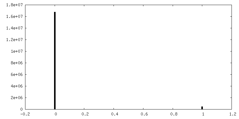
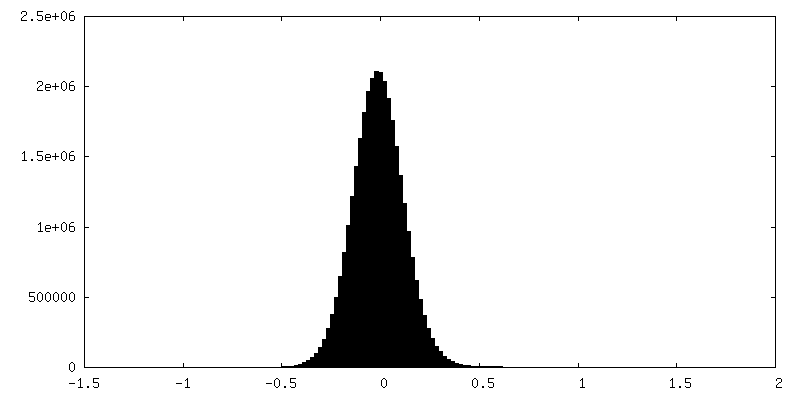
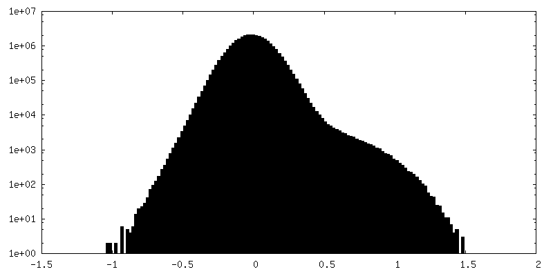
-Mask #1
| File | emd_22879_msk_1.map | ||||||||||||
|---|---|---|---|---|---|---|---|---|---|---|---|---|---|
| Projections & Slices |
| ||||||||||||
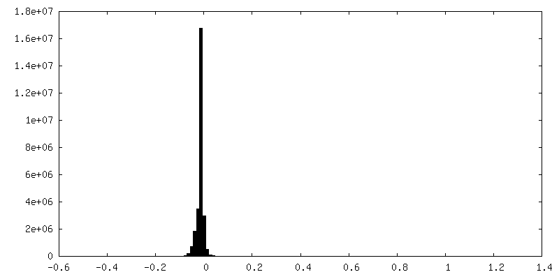
| Density Histograms |


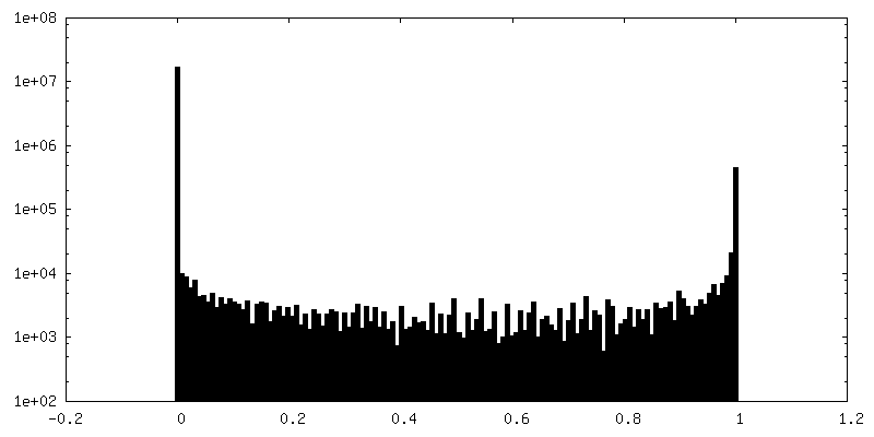
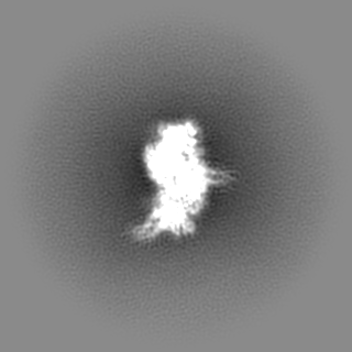
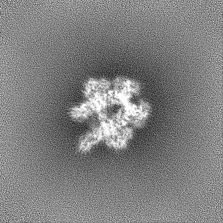
-Additional map: Unsharpened map
| File | emd_22879_additional_1.map | ||||||||||||
|---|---|---|---|---|---|---|---|---|---|---|---|---|---|
| Annotation | Unsharpened map | ||||||||||||
| Projections & Slices |
| ||||||||||||
| Density Histograms |


-Half map: Half map A
| File | emd_22879_half_map_1.map | ||||||||||||
|---|---|---|---|---|---|---|---|---|---|---|---|---|---|
| Annotation | Half map A | ||||||||||||
| Projections & Slices |
| ||||||||||||
| Density Histograms |


-Half map: Half map B
| File | emd_22879_half_map_2.map | ||||||||||||
|---|---|---|---|---|---|---|---|---|---|---|---|---|---|
| Annotation | Half map B | ||||||||||||
| Projections & Slices |
| ||||||||||||
| Density Histograms |


- Sample components
-Entire : MDB1 Fab
| Entire | Name: MDB1 Fab |
|---|---|
| Components |
|
-Supramolecule #1: MDB1 Fab
| Supramolecule | Name: MDB1 Fab / type: complex / ID: 1 / Parent: 0 / Macromolecule list: #1-#3 |
|---|---|
| Source (natural) | Organism: Homo sapiens (human) |
-Supramolecule #2: MDB1 Fab in complex with RIFIN
| Supramolecule | Name: MDB1 Fab in complex with RIFIN / type: complex / ID: 2 / Parent: 1 / Macromolecule list: #1-#3 |
|---|---|
| Source (natural) | Organism: Homo sapiens (human) |
-Macromolecule #1: MDB1 Fab heavy chain
| Macromolecule | Name: MDB1 Fab heavy chain / type: protein_or_peptide / ID: 1 / Number of copies: 2 / Enantiomer: LEVO |
|---|---|
| Source (natural) | Organism: Homo sapiens (human) |
| Molecular weight | Theoretical: 46.384973 KDa |
| Recombinant expression | Organism: Homo sapiens (human) |
| Sequence | String: QITLKESGPT LVERTQTLTL TCTFSGFSLT TDGLGVGWIR QPPGKALEWL ALIYYDDVRR YRPSLKSRLT ITKDTSKNRV VLTVTDMDP VDAGTYFCVH ASLFNLGSRN VYFRYWGQGT LVSVSSGVSK KPSLSVQPGP IVAPEETLTL QCGSDAGYNR F VLYKDGER ...String: QITLKESGPT LVERTQTLTL TCTFSGFSLT TDGLGVGWIR QPPGKALEWL ALIYYDDVRR YRPSLKSRLT ITKDTSKNRV VLTVTDMDP VDAGTYFCVH ASLFNLGSRN VYFRYWGQGT LVSVSSGVSK KPSLSVQPGP IVAPEETLTL QCGSDAGYNR F VLYKDGER DFLQLAGAQP QAGLSQANFT LGPVSRSYGG QYRCYGAHNL SSEWSAPSDP LDILIAGQFY DRVSLSVQPG PT VASGENV TLLCQSQGWM QTFLLTKEGA ADDPWRLRST YQSQKYQAEF PMGPVTSAHA GTYRCYGSQS SKPYLLTHPS DPL ELVVSA STKGPSVFPL APCSRSTSES TAALGCLVKD YFPEPVTVSW NSGALTSGVH TFPAVLQSSG LYSLSSVVTV PSSS LGTKT YTCNVDHKPS NTKVDKRVES KY |
-Macromolecule #2: MDB1 Fab light chain
| Macromolecule | Name: MDB1 Fab light chain / type: protein_or_peptide / ID: 2 / Number of copies: 2 / Enantiomer: LEVO |
|---|---|
| Source (natural) | Organism: Homo sapiens (human) |
| Molecular weight | Theoretical: 22.870232 KDa |
| Recombinant expression | Organism: Homo sapiens (human) |
| Sequence | String: SYDLTQPPSV SVSPGQTARI TCSGDSLARQ FASWYQQKPG QSPVLLIYKN IERLSAIPER FSGSTSGTSV TLTISGVQAE DEADYFCQS ADDSGSYSLF GTGTRVSVLG QPKANPTVTL FPPSSEELQA NKATLVCLIS DFYPGAVTVA WKADGSPVKA G VETTKPSK ...String: SYDLTQPPSV SVSPGQTARI TCSGDSLARQ FASWYQQKPG QSPVLLIYKN IERLSAIPER FSGSTSGTSV TLTISGVQAE DEADYFCQS ADDSGSYSLF GTGTRVSVLG QPKANPTVTL FPPSSEELQA NKATLVCLIS DFYPGAVTVA WKADGSPVKA G VETTKPSK QSNNKYAASS YLSLTPEQWK SHRSYSCQVT HEGSTVEKTV APTECS |
-Macromolecule #3: Rifin
| Macromolecule | Name: Rifin / type: protein_or_peptide / ID: 3 / Number of copies: 2 / Enantiomer: LEVO |
|---|---|
| Source (natural) | Organism: |
| Molecular weight | Theoretical: 15.420644 KDa |
| Recombinant expression | Organism: Homo sapiens (human) |
| Sequence | String: KELAEKAGAL AGEAARIPAA IDAVIEGIKS KFSIDTLGGE ALKSVIDGTN YYDASYITTA IYNKFQVSSC LPSVPFLGGP PVPGAGANK PICSAVDKLY LGSGNFLDKS SLPGSIQKDV AKIVAGAEQA AKAKAAMVAS DKTLAVETAK KQ UniProtKB: Rifin |
-Macromolecule #5: 2-acetamido-2-deoxy-beta-D-glucopyranose
| Macromolecule | Name: 2-acetamido-2-deoxy-beta-D-glucopyranose / type: ligand / ID: 5 / Number of copies: 2 / Formula: NAG |
|---|---|
| Molecular weight | Theoretical: 221.208 Da |
| Chemical component information |  ChemComp-NAG: |
-Experimental details
-Structure determination
| Method | cryo EM |
|---|---|
 Processing Processing | single particle reconstruction |
| Aggregation state | particle |
-Sample preparation
| Concentration | 1 mg/mL |
|---|---|
| Buffer | pH: 7.4 / Component - Formula: PBS |
| Grid | Model: C-flat-1.2/1.3 / Material: COPPER / Support film - Material: CARBON / Support film - topology: HOLEY / Pretreatment - Type: PLASMA CLEANING |
| Vitrification | Cryogen name: ETHANE / Chamber humidity: 90 % / Chamber temperature: 293 K / Instrument: FEI VITROBOT MARK IV |
- Electron microscopy
| Microscope | FEI TITAN KRIOS |
|---|---|
| Image recording | Film or detector model: GATAN K3 (6k x 4k) / Number grids imaged: 1 / Number real images: 6473 / Average exposure time: 2.0 sec. / Average electron dose: 70.72 e/Å2 |
| Electron beam | Acceleration voltage: 300 kV / Electron source:  FIELD EMISSION GUN FIELD EMISSION GUN |
| Electron optics | C2 aperture diameter: 70.0 µm / Illumination mode: FLOOD BEAM / Imaging mode: BRIGHT FIELD |
| Sample stage | Specimen holder model: FEI TITAN KRIOS AUTOGRID HOLDER / Cooling holder cryogen: NITROGEN |
| Experimental equipment |  Model: Titan Krios / Image courtesy: FEI Company |
+Image processing
-Atomic model buiding 1
| Initial model | PDB ID: Chain - Chain ID: A / Chain - Source name: PDB / Chain - Initial model type: experimental model |
|---|---|
| Refinement | Space: REAL / Protocol: FLEXIBLE FIT |
| Output model | PDB-7khf: |

