 Movie
Movie Controller

[English] 日本語
 Yorodumi
- PDB-7kfk: Crystal structure of LILRB1 D3D4 domain in complex with Plasmodiu... -
+ Open data

- Basic information
| Entry | Database: PDB / ID: 7kfk | ||||||
|---|---|---|---|---|---|---|---|
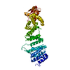
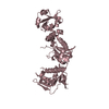
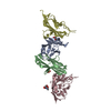
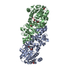
| Title | Crystal structure of LILRB1 D3D4 domain in complex with Plasmodium RIFIN (PF3D7_1373400) V2 domain | ||||||
 Components Components |
| ||||||
 Keywords Keywords | IMMUNE SYSTEM / LILRB1 / RIFIN / Malaria | ||||||
| Function / homology | Variant surface antigen Rifin / Rifin / Rifin / Isoform 2 of Leukocyte immunoglobulin-like receptor subfamily B member 1 Function and homology information Function and homology information | ||||||
| Biological species |  Homo sapiens (human) Homo sapiens (human) | ||||||
| Method |  X-RAY DIFFRACTION / SYNCHROTRON / MOLECULAR REPLACEMENT / Resolution: 2.63 Å X-RAY DIFFRACTION / SYNCHROTRON / MOLECULAR REPLACEMENT / Resolution: 2.63 Å | ||||||
 Authors Authors | Xu, K. / Kwong, P.D. | ||||||
 Citation Citation | Journal: Nature / Year: 2021 Title: Structural basis of malaria RIFIN binding by LILRB1-containing antibodies. Authors: Yiwei Chen / Kai Xu / Luca Piccoli / Mathilde Foglierini / Joshua Tan / Wenjie Jin / Jason Gorman / Yaroslav Tsybovsky / Baoshan Zhang / Boubacar Traore / Chiara Silacci-Fregni / Claudia ...Authors: Yiwei Chen / Kai Xu / Luca Piccoli / Mathilde Foglierini / Joshua Tan / Wenjie Jin / Jason Gorman / Yaroslav Tsybovsky / Baoshan Zhang / Boubacar Traore / Chiara Silacci-Fregni / Claudia Daubenberger / Peter D Crompton / Roger Geiger / Federica Sallusto / Peter D Kwong / Antonio Lanzavecchia /   Abstract: Some Plasmodium falciparum repetitive interspersed families of polypeptides (RIFINs)-variant surface antigens that are expressed on infected erythrocytes-bind to the inhibitory receptor LAIR1, and ...Some Plasmodium falciparum repetitive interspersed families of polypeptides (RIFINs)-variant surface antigens that are expressed on infected erythrocytes-bind to the inhibitory receptor LAIR1, and insertion of DNA that encodes LAIR1 into immunoglobulin genes generates RIFIN-specific antibodies. Here we address the general relevance of this finding by searching for antibodies that incorporate LILRB1, another inhibitory receptor that binds to β2 microglobulin and RIFINs through their apical domains. By screening plasma from a cohort of donors from Mali, we identified individuals with LILRB1-containing antibodies. B cell clones isolated from three donors showed large DNA insertions in the switch region that encodes non-apical LILRB1 extracellular domain 3 and 4 (D3D4) or D3 alone in the variable-constant (VH-CH1) elbow. Through mass spectrometry and binding assays, we identified a large set of RIFINs that bind to LILRB1 D3. Crystal and cryo-electron microscopy structures of a RIFIN in complex with either LILRB1 D3D4 or a D3D4-containing antibody Fab revealed a mode of RIFIN-LILRB1 D3 interaction that is similar to that of RIFIN-LAIR1. The Fab showed an unconventional triangular architecture with the inserted LILRB1 domains opening up the VH-CH1 elbow without affecting VH-VL or CH1-CL pairing. Collectively, these findings show that RIFINs bind to LILRB1 through D3 and illustrate, with a naturally selected example, the general principle of creating novel antibodies by inserting receptor domains into the VH-CH1 elbow. | ||||||
| History |
|
- Structure visualization
| Structure viewer | Molecule: MolmilJmol/JSmol |
|---|
- Downloads & links
-Download
| PDBx/mmCIF format | 7kfk.cif.gz | 144.5 KB | Display | PDBx/mmCIF format |
|---|---|---|---|---|
| PDB format | pdb7kfk.ent.gz | 110.5 KB | Display | PDB format |
| PDBx/mmJSON format | 7kfk.json.gz | Tree view | PDBx/mmJSON format | |
| Others |  Other downloads Other downloads |
-Validation report
| Arichive directory | https://data.pdbj.org/pub/pdb/validation_reports/kf/7kfkftp://data.pdbj.org/pub/pdb/validation_reports/kf/7kfk | HTTPS FTP |
|---|
-Related structure data
| Related structure data |  7khfC  4llaS S: Starting model for refinement C: citing same article ( |
|---|---|
| Similar structure data |


-Links
 PDBj
PDBj- Assembly
| Deposited unit | 
| |||||||||||||||||||||||||||||||||||||||||||||||||||||||||||||||||||||||||||||||
|---|---|---|---|---|---|---|---|---|---|---|---|---|---|---|---|---|---|---|---|---|---|---|---|---|---|---|---|---|---|---|---|---|---|---|---|---|---|---|---|---|---|---|---|---|---|---|---|---|---|---|---|---|---|---|---|---|---|---|---|---|---|---|---|---|---|---|---|---|---|---|---|---|---|---|---|---|---|---|---|---|
| 1 | 
| |||||||||||||||||||||||||||||||||||||||||||||||||||||||||||||||||||||||||||||||
| 2 | 
| |||||||||||||||||||||||||||||||||||||||||||||||||||||||||||||||||||||||||||||||
| Unit cell |
| |||||||||||||||||||||||||||||||||||||||||||||||||||||||||||||||||||||||||||||||
| Noncrystallographic symmetry (NCS) | NCS domain:
NCS domain segments:
|