 Movie
Movie Controller


+ Open data

- Basic information
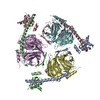
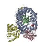
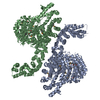
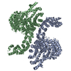
| Entry | Database: PDB / ID: 1mhs | ||||||
|---|---|---|---|---|---|---|---|
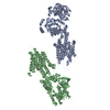
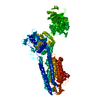
| Title | Model of Neurospora crassa proton ATPase | ||||||
 Components Components | Plasma Membrane ATPase | ||||||
 Keywords Keywords | MEMBRANE PROTEIN / PROTON TRANSPORT / ion transport / proton pump / P-type ATPase / active transport | ||||||
| Function / homology |  Function and homology information Function and homology informationP-type H+-exporting transporter / proton export across plasma membrane / P-type proton-exporting transporter activity / proton transmembrane transport / regulation of intracellular pH / ATP hydrolysis activity / ATP binding / metal ion binding / plasma membrane Similarity search - Function | ||||||
| Biological species |  Neurospora crassa (fungus) Neurospora crassa (fungus) | ||||||
| Method | ELECTRON CRYSTALLOGRAPHY / electron crystallography / cryo EM / Resolution: 8 Å | ||||||
 Authors Authors | Kuhlbrandt, W. | ||||||
 Citation Citation | Journal: Science / Year: 2002 Title: Structure, mechanism, and regulation of the Neurospora plasma membrane H+-ATPase. Authors: Werner Kühlbrandt / Johan Zeelen / Jens Dietrich /  Abstract: Proton pumps in the plasma membrane of plants and yeasts maintain the intracellular pH and membrane potential. To gain insight into the molecular mechanisms of proton pumping, we built an atomic ...Proton pumps in the plasma membrane of plants and yeasts maintain the intracellular pH and membrane potential. To gain insight into the molecular mechanisms of proton pumping, we built an atomic homology model of the proton pump based on the 2.6 angstrom x-ray structure of the related Ca2+ pump from rabbit sarcoplasmic reticulum. The model, when fitted to an 8 angstrom map of the Neurospora proton pump determined by electron microscopy, reveals the likely path of the proton through the membrane and shows that the nucleotide-binding domain rotates by approximately 70 degrees to deliver adenosine triphosphate (ATP) to the phosphorylation site. A synthetic peptide corresponding to the carboxyl-terminal regulatory domain stimulates ATPase activity, suggesting a mechanism for proton transport regulation. #1: Journal: Nature / Year: 1998Title: Three-dimensional map of the plasma membrane H+-ATPase in the open conformation Authors: AUER, M. / SCARBOROUGH, G.A. / KUHLBRANDT, W. | ||||||
| History |
| ||||||
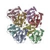
| Remark 295 | NON-CRYSTALLOGRAPHIC SYMMETRY THE TRANSFORMATIONS PRESENTED ON THE MTRIX RECORDS BELOW DESCRIBE ... NON-CRYSTALLOGRAPHIC SYMMETRY THE TRANSFORMATIONS PRESENTED ON THE MTRIX RECORDS BELOW DESCRIBE NON-CRYSTALLOGRAPHIC RELATIONSHIPS AMONG ATOMS IN THIS ENTRY. APPLYING THE APPROPRIATE MTRIX TRANSFORMATION TO THE RESIDUES LISTED FIRST WILL YIELD APPROXIMATE COORDINATES FOR THE RESIDUES LISTED SECOND. CHAIN IDENTIFIERS GIVEN AS "?" REFER TO CHAINS FOR WHICH ATOMS ARE NOT FOUND IN THE ENTRY. APPLIED TO TRANSFORMED TO TRANSFORM CHAIN RESIDUES CHAIN RESIDUES RMSD SSS M 1 A 1 .. 920 B 1 .. 920 ? WHERE SSS -> COLUMNS 8-10 OF MTRIX RECORDS | ||||||
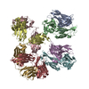
| Remark 300 | BIOMOLECULE: 1 THIS ENTRY CONTAINS A PORTION OF THE BIOLOGICALLY SIGNIFICANT MULTIMER. SEE REMARK ...BIOMOLECULE: 1 THIS ENTRY CONTAINS A PORTION OF THE BIOLOGICALLY SIGNIFICANT MULTIMER. SEE REMARK 350 FOR INFORMATION ON GENERATING THE BIOLOGICAL MOLECULE(S) ASSEMBLY COMPONENTS COM_ID: 1 NAME:PLASMA MEMBRANE PROTON ATPASE HEXAMERIC ASSEMBLY |
- Structure visualization
| Movie |
Movie viewer |
|---|---|
| Structure viewer | Molecule: MolmilJmol/JSmol |
- Downloads & links
-Download
| PDBx/mmCIF format | 1mhs.cif.gz | 317.1 KB | Display | PDBx/mmCIF format |
|---|---|---|---|---|
| PDB format | pdb1mhs.ent.gz | 249.7 KB | Display | PDB format |
| PDBx/mmJSON format | 1mhs.json.gz | Tree view | PDBx/mmJSON format | |
| Others |  Other downloads Other downloads |
-Validation report
| Arichive directory | https://data.pdbj.org/pub/pdb/validation_reports/mh/1mhsftp://data.pdbj.org/pub/pdb/validation_reports/mh/1mhs | HTTPS FTP |
|---|
-Related structure data
| Related structure data | |
|---|---|
| Similar structure data |


-Links
 PDBj
PDBj


- Assembly
| Deposited unit | 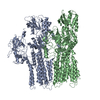
| ||||||||
|---|---|---|---|---|---|---|---|---|---|
| 1 |
| ||||||||
| Unit cell |
| ||||||||
| Noncrystallographic symmetry (NCS) | NCS oper: (Code: given / Matrix: (0.5, 0.866), |
-Components
| #1: Protein | Mass: 99984.359 Da / Num. of mol.: 2 / Source method: isolated from a natural source Details: isolated from plasma membrane of cultured Neurospora cells Source: (natural) Neurospora crassa (fungus) / Strain: FGSC 4761 / References: UniProt: P07038, EC: 3.6.3.6 |
|---|
-Experimental details
-Experiment
| Experiment | Method: ELECTRON CRYSTALLOGRAPHY |
|---|---|
| EM experiment | Aggregation state: 2D ARRAY / 3D reconstruction method: electron crystallography |
- Sample preparation
| Component |
| |||||||||||||||
|---|---|---|---|---|---|---|---|---|---|---|---|---|---|---|---|---|
| Buffer solution | Name: 100 mM ammonium sulphate, Tris-HCl, 30% glycerol, 10.5% PEG 4000 pH: 6.8 Details: 100 mM ammonium sulphate, Tris-HCl, 30% glycerol, 10.5% PEG 4000 | |||||||||||||||
| Specimen | Conc.: 1 mg/ml / Embedding applied: NO / Shadowing applied: NO / Staining applied: NO / Vitrification applied: YES | |||||||||||||||
| Specimen support | Details: hydrophobic carbon support film on 400 mesh gold-plated Cu EM grid | |||||||||||||||
| Vitrification | Details: 2D crystals were vitrified by immersion in liquid nitrogen | |||||||||||||||
| Crystal grow | *PLUS Method: other / Details: MRC ELECTRON CRYSTALLOGRAPHY |
-Data collection
| Microscopy | Model: JEOL 3000SFF Details: Images were also recorded on JEOL 2000 EX and Philips CM200 FEG electron microscopes at 80 K at a dose of 1000 e/nm**2 |
|---|---|
| Electron gun | Electron source:  FIELD EMISSION GUN / Accelerating voltage: 300 kV / Illumination mode: FLOOD BEAM FIELD EMISSION GUN / Accelerating voltage: 300 kV / Illumination mode: FLOOD BEAM |
| Electron lens | Mode: BRIGHT FIELD / Nominal magnification: 50000 X / Calibrated magnification: 50000 X / Nominal defocus max: 1200 nm / Nominal defocus min: 600 nm / Cs: 1.6 mm |
| Specimen holder | Temperature: 4 K / Tilt angle max: 60 ° / Tilt angle min: 0 ° |
| Image recording | Electron dose: 25 e/Å2 / Film or detector model: KODAK SO-163 FILM |
| Image scans | Num. digital images: 60 |
| Radiation | Scattering type: electron |
| Radiation wavelength | Relative weight: 1 |
- Processing
| EM software | Name: MRC / Category: 3D reconstruction | ||||||||||||
|---|---|---|---|---|---|---|---|---|---|---|---|---|---|
| CTF correction | Details: crystallographic | ||||||||||||
| 3D reconstruction | Resolution: 8 Å / Nominal pixel size: 1.4 Å / Actual pixel size: 1.4 Å Magnification calibration: electron diffraction of standard specimen Symmetry type: 2D CRYSTAL | ||||||||||||
| Atomic model building | Protocol: OTHER / Space: REAL / Target criteria: best visual fit using the program o / Details: METHOD--manual REFINEMENT PROTOCOL--manual fit | ||||||||||||
| Atomic model building | Details: this entry | ||||||||||||
| Refinement | Highest resolution: 8 Å | ||||||||||||
| Refinement step | Cycle: LAST / Highest resolution: 8 Å
|
