 Movie
Movie Controller

[English] 日本語
 Yorodumi
- PDB-4o6h: 2.8A crystal structure of Lymphocytic Choriomeningitis Virus Nucl... -
+ Open data

- Basic information
| Entry | Database: PDB / ID: 4o6h | ||||||
|---|---|---|---|---|---|---|---|
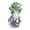
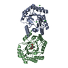
| Title | 2.8A crystal structure of Lymphocytic Choriomeningitis Virus Nucleoprotein C-terminal Domain | ||||||
 Components Components | Nucleoprotein | ||||||
 Keywords Keywords | HYDROLASE / Exoribonuclease | ||||||
| Function / homology |  Function and homology information Function and homology informationsymbiont-mediated suppression of host cytoplasmic pattern recognition receptor signaling pathway via inhibition of IKBKE activity / RNA-templated viral transcription / negative stranded viral RNA replication / helical viral capsid / viral nucleocapsid / Hydrolases; Acting on ester bonds; Exoribonucleases producing 5'-phosphomonoesters / symbiont-mediated suppression of host NF-kappaB cascade / host cell cytoplasm / ribonucleoprotein complex / hydrolase activity ...symbiont-mediated suppression of host cytoplasmic pattern recognition receptor signaling pathway via inhibition of IKBKE activity / RNA-templated viral transcription / negative stranded viral RNA replication / helical viral capsid / viral nucleocapsid / Hydrolases; Acting on ester bonds; Exoribonucleases producing 5'-phosphomonoesters / symbiont-mediated suppression of host NF-kappaB cascade / host cell cytoplasm / ribonucleoprotein complex / hydrolase activity / RNA binding / metal ion binding Similarity search - Function | ||||||
| Biological species |  Lymphocytic choriomeningitis virus Lymphocytic choriomeningitis virus | ||||||
| Method |  X-RAY DIFFRACTION / SYNCHROTRON / MOLECULAR REPLACEMENT / Resolution: 2.8 Å X-RAY DIFFRACTION / SYNCHROTRON / MOLECULAR REPLACEMENT / Resolution: 2.8 Å | ||||||
 Authors Authors | West, B.R. / Hastie, K.M. / Saphire, E.O. | ||||||
 Citation Citation | Journal: Acta Crystallogr.,Sect.D / Year: 2014 Title: Structure of the LCMV nucleoprotein provides a template for understanding arenavirus replication and immunosuppression. Authors: West, B.R. / Hastie, K.M. / Saphire, E.O. | ||||||
| History |
|
- Structure visualization
| Structure viewer | Molecule: MolmilJmol/JSmol |
|---|
- Downloads & links
-Download
| PDBx/mmCIF format | 4o6h.cif.gz | 577.2 KB | Display | PDBx/mmCIF format |
|---|---|---|---|---|
| PDB format | pdb4o6h.ent.gz | 481.9 KB | Display | PDB format |
| PDBx/mmJSON format | 4o6h.json.gz | Tree view | PDBx/mmJSON format | |
| Others |  Other downloads Other downloads |
-Validation report
| Arichive directory | https://data.pdbj.org/pub/pdb/validation_reports/o6/4o6hftp://data.pdbj.org/pub/pdb/validation_reports/o6/4o6h | HTTPS FTP |
|---|
-Related structure data
| Related structure data | 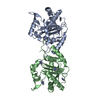 4o6iC 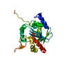 3q7cS C: citing same article ( S: Starting model for refinement |
|---|---|
| Similar structure data |


-Links
 PDBj
PDBj


- Assembly
| Deposited unit | 
| ||||||||
|---|---|---|---|---|---|---|---|---|---|
| 1 | 
| ||||||||
| 2 | 
| ||||||||
| 3 | 
| ||||||||
| 4 | 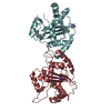
| ||||||||
| Unit cell |
|
-Components
| #1: Protein | Mass: 26400.145 Da / Num. of mol.: 8 / Fragment: C-Terminal Domain Source method: isolated from a genetically manipulated source Source: (gene. exp.) Lymphocytic choriomeningitis virus / Strain: Armstrong / Gene: N / Plasmid: pET46 / Production host:  #2: Chemical | ChemComp-ZN /   Mass: 65.409 Da / Num. of mol.: 8 / Source method: obtained synthetically / Formula: Zn Mass: 65.409 Da / Num. of mol.: 8 / Source method: obtained synthetically / Formula: Zn#3: Chemical | ChemComp-IMD /   Mass: 69.085 Da / Num. of mol.: 4 / Source method: obtained synthetically / Formula: C3H5N2 Mass: 69.085 Da / Num. of mol.: 4 / Source method: obtained synthetically / Formula: C3H5N2#4: Chemical | ChemComp-MG /   Mass: 24.305 Da / Num. of mol.: 4 / Source method: obtained synthetically / Formula: Mg Mass: 24.305 Da / Num. of mol.: 4 / Source method: obtained synthetically / Formula: Mg#5: Chemical | ChemComp-K /   Mass: 39.098 Da / Num. of mol.: 4 / Source method: obtained synthetically / Formula: K Mass: 39.098 Da / Num. of mol.: 4 / Source method: obtained synthetically / Formula: K |
|---|
-Experimental details
-Experiment
| Experiment | Method: X-RAY DIFFRACTION / Number of used crystals: 1 |
|---|
- Sample preparation
| Crystal | Density Matthews: 2.94 Å3/Da / Density % sol: 58.2 % |
|---|---|
| Crystal grow | Temperature: 295 K / Method: vapor diffusion, sitting drop / pH: 8 Details: 0.35M potassium phosphate, 20% PEG 3350, cryoprotected with 20% glycerol, pH 8, VAPOR DIFFUSION, SITTING DROP, temperature 295K |
-Data collection
| Diffraction | Mean temperature: 100 K |
|---|---|
| Diffraction source | Source: SYNCHROTRON / Site: ALS  / Beamline: 8.2.2 / Wavelength: 0.9793 Å / Beamline: 8.2.2 / Wavelength: 0.9793 Å |
| Detector | Type: ADSC QUANTUM 315r / Detector: CCD / Date: Oct 18, 2012 |
| Radiation | Monochromator: Double crystal, Si(111) / Protocol: SINGLE WAVELENGTH / Monochromatic (M) / Laue (L): M / Scattering type: x-ray |
| Radiation wavelength | Wavelength: 0.9793 Å / Relative weight: 1 |
| Reflection | Resolution: 2.8→47.264 Å / Num. all: 60576 / Num. obs: 58941 / % possible obs: 97.3 % / Observed criterion σ(F): 1.6 / Observed criterion σ(I): 1.6 |
- Processing
| Software |
| ||||||||||||||||||||||||||||||||||||||||||||||||||||||||||||||||||||||||||||||||||||||||||||||||||||||||||||||||||||||||||||||||||||||||||||||||||||||||||
|---|---|---|---|---|---|---|---|---|---|---|---|---|---|---|---|---|---|---|---|---|---|---|---|---|---|---|---|---|---|---|---|---|---|---|---|---|---|---|---|---|---|---|---|---|---|---|---|---|---|---|---|---|---|---|---|---|---|---|---|---|---|---|---|---|---|---|---|---|---|---|---|---|---|---|---|---|---|---|---|---|---|---|---|---|---|---|---|---|---|---|---|---|---|---|---|---|---|---|---|---|---|---|---|---|---|---|---|---|---|---|---|---|---|---|---|---|---|---|---|---|---|---|---|---|---|---|---|---|---|---|---|---|---|---|---|---|---|---|---|---|---|---|---|---|---|---|---|---|---|---|---|---|---|---|---|
| Refinement | Method to determine structure: MOLECULAR REPLACEMENT Starting model: PDB ENTRY 3Q7C Resolution: 2.8→47.264 Å / SU ML: 0.46 / σ(F): 1.36 / Phase error: 35.34 / Stereochemistry target values: ML
| ||||||||||||||||||||||||||||||||||||||||||||||||||||||||||||||||||||||||||||||||||||||||||||||||||||||||||||||||||||||||||||||||||||||||||||||||||||||||||
| Solvent computation | Shrinkage radii: 0.9 Å / VDW probe radii: 1.11 Å / Solvent model: FLAT BULK SOLVENT MODEL | ||||||||||||||||||||||||||||||||||||||||||||||||||||||||||||||||||||||||||||||||||||||||||||||||||||||||||||||||||||||||||||||||||||||||||||||||||||||||||
| Refinement step | Cycle: LAST / Resolution: 2.8→47.264 Å
| ||||||||||||||||||||||||||||||||||||||||||||||||||||||||||||||||||||||||||||||||||||||||||||||||||||||||||||||||||||||||||||||||||||||||||||||||||||||||||
| Refine LS restraints |
| ||||||||||||||||||||||||||||||||||||||||||||||||||||||||||||||||||||||||||||||||||||||||||||||||||||||||||||||||||||||||||||||||||||||||||||||||||||||||||
| LS refinement shell |
|
