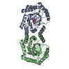
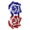
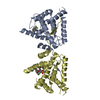
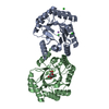
Entry Database : PDB / ID : 6ku0Title Crystal structure of MyoVa-GTD in complex with MICAL1-GTBM Peptide from [F-actin]-monooxygenase MICAL1 Unconventional myosin-Va Keywords / / Function / homology Function Domain/homology Component
/ / / / / / / / / / / / / / / / / / / / / / / / / / / / / / / / / / / / / / / / / / / / / / / / / / / / / / / / / / / / / / / / / / / / / / / / / / / / / / / / / / / / / / / / / / / / / / / / / / / / / / / / / / / / / / / / / / / / / / / / / / / / / / / / / / / / / / / / / / / / / / / / / / / / / / / / / / / / Biological species Mus musculus (house mouse)Homo sapiens (human)Method / / / Resolution : 1.6 Å Authors Niu, F. / Wei, Z. Funding support Organization Grant number Country National Natural Science Foundation of China 31800643 National Natural Science Foundation of China 31770791 National Natural Science Foundation of China 31570741
Journal : Sci Adv / Year : 2020Title : F-actin disassembly factor MICAL1 binding to Myosin Va mediates cargo unloading during cytokinesis.Authors : Niu, F. / Sun, K. / Wei, W. / Yu, C. / Wei, Z. History Deposition Aug 29, 2019 Deposition site / Processing site Revision 1.0 Sep 2, 2020 Provider / Type Revision 1.1 Nov 18, 2020 Group / Category / citation_authorItem _citation.country / _citation.journal_abbrev ... _citation.country / _citation.journal_abbrev / _citation.journal_id_CSD / _citation.journal_id_ISSN / _citation.journal_volume / _citation.pdbx_database_id_DOI / _citation.pdbx_database_id_PubMed / _citation.title / _citation.year / _citation_author.identifier_ORCID Revision 1.2 Nov 22, 2023 Group / Database references / Refinement descriptionCategory chem_comp_atom / chem_comp_bond ... chem_comp_atom / chem_comp_bond / database_2 / pdbx_initial_refinement_model Item / _database_2.pdbx_database_accession
Show all Show less
 Movie
Movie Controller


 Open data
 Basic information
 Components
 Keywords
 Function and homology information
 Homo sapiens (human)
 X-RAY DIFFRACTION /
  Authors
 China, 3items
  Citation
 Structure visualization
 Downloads & links
 Other downloads
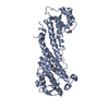


 PDBj
PDBj


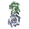
 Assembly


 Mass: 62.068 Da / Num. of mol.: 5 / Source method: obtained synthetically / Formula: C2H6O2
 Mass: 18.015 Da / Num. of mol.: 700 / Source method: isolated from a natural source / Formula: H2O
 Sample preparation
 Processing