Movie
Movie Controller

[English] 日本語
 Yorodumi
- PDB-7k48: Structure of NavAb/Nav1.7-VS2A chimera trapped in the resting sta... -
+ Open data

- Basic information
| Entry | Database: PDB / ID: 7k48 | |||||||||||||||
|---|---|---|---|---|---|---|---|---|---|---|---|---|---|---|---|---|
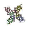
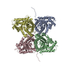
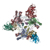
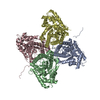
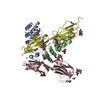
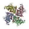
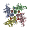
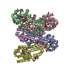
| Title | Structure of NavAb/Nav1.7-VS2A chimera trapped in the resting state by tarantula toxin m3-Huwentoxin-IV | |||||||||||||||
 Components Components |
| |||||||||||||||
 Keywords Keywords | MEMBRANE PROTEIN / Ion channel / Voltage-gated sodium channel / Gating-modifier toxin | |||||||||||||||
| Function / homology |  Function and homology information Function and homology informationaction potential propagation / detection of mechanical stimulus involved in sensory perception / host cell presynaptic membrane / cardiac muscle cell action potential involved in contraction / voltage-gated sodium channel complex / node of Ranvier / voltage-gated sodium channel activity / Interaction between L1 and Ankyrins / detection of maltose stimulus / ion channel inhibitor activity ...action potential propagation / detection of mechanical stimulus involved in sensory perception / host cell presynaptic membrane / cardiac muscle cell action potential involved in contraction / voltage-gated sodium channel complex / node of Ranvier / voltage-gated sodium channel activity / Interaction between L1 and Ankyrins / detection of maltose stimulus / ion channel inhibitor activity / maltose transport complex / Phase 0 - rapid depolarisation / detection of temperature stimulus involved in sensory perception of pain / carbohydrate transport / behavioral response to pain / sodium channel regulator activity / carbohydrate transmembrane transporter activity / maltose binding / maltose transport / maltodextrin transmembrane transport / neuronal action potential / ATP-binding cassette (ABC) transporter complex, substrate-binding subunit-containing / sensory perception of pain / ATP-binding cassette (ABC) transporter complex / axon terminus / post-embryonic development / sodium ion transmembrane transport / cell chemotaxis / circadian rhythm / response to toxic substance / Sensory perception of sweet, bitter, and umami (glutamate) taste / toxin activity / outer membrane-bounded periplasmic space / periplasmic space / inflammatory response / axon / DNA damage response / extracellular region / membrane / metal ion binding / identical protein binding / plasma membrane Similarity search - Function | |||||||||||||||
| Biological species |  Arcobacter butzleri (bacteria) Arcobacter butzleri (bacteria) Homo sapiens (human) Homo sapiens (human) Haplopelma schmidti (Chinese earth tiger) Haplopelma schmidti (Chinese earth tiger) | |||||||||||||||
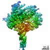
| Method | ELECTRON MICROSCOPY / single particle reconstruction / cryo EM / Resolution: 3.6 Å | |||||||||||||||
 Authors Authors | Wisedchaisri, G. / Tonggu, L. / Gamal El-Din, T.M. / McCord, E. / Zheng, N. / Catterall, W.A. | |||||||||||||||
| Funding support |  United States, 4items United States, 4items
| |||||||||||||||
 Citation Citation | Journal: Mol Cell / Year: 2021 Title: Structural Basis for High-Affinity Trapping of the Na1.7 Channel in Its Resting State by Tarantula Toxin. Authors: Goragot Wisedchaisri / Lige Tonggu / Tamer M Gamal El-Din / Eedann McCord / Ning Zheng / William A Catterall / Abstract: Voltage-gated sodium channels initiate electrical signals and are frequently targeted by deadly gating-modifier neurotoxins, including tarantula toxins, which trap the voltage sensor in its resting ...Voltage-gated sodium channels initiate electrical signals and are frequently targeted by deadly gating-modifier neurotoxins, including tarantula toxins, which trap the voltage sensor in its resting state. The structural basis for tarantula-toxin action remains elusive because of the difficulty of capturing the functionally relevant form of the toxin-channel complex. Here, we engineered the model sodium channel NaAb with voltage-shifting mutations and the toxin-binding site of human Na1.7, an attractive pain target. This mutant chimera enabled us to determine the cryoelectron microscopy (cryo-EM) structure of the channel functionally arrested by tarantula toxin. Our structure reveals a high-affinity resting-state-specific toxin-channel interaction between a key lysine residue that serves as a "stinger" and penetrates a triad of carboxyl groups in the S3-S4 linker of the voltage sensor. By unveiling this high-affinity binding mode, our studies establish a high-resolution channel-docking and resting-state locking mechanism for huwentoxin-IV and provide guidance for developing future resting-state-targeted analgesic drugs. | |||||||||||||||
| History |
|
- Structure visualization
| Movie |
Movie viewer |
|---|---|
| Structure viewer | Molecule: MolmilJmol/JSmol |
- Downloads & links
-Download
| PDBx/mmCIF format | 7k48.cif.gz | 214.8 KB | Display | PDBx/mmCIF format |
|---|---|---|---|---|
| PDB format | pdb7k48.ent.gz | 160.6 KB | Display | PDB format |
| PDBx/mmJSON format | 7k48.json.gz | Tree view | PDBx/mmJSON format | |
| Others |  Other downloads Other downloads |
-Validation report
| Arichive directory | https://data.pdbj.org/pub/pdb/validation_reports/k4/7k48ftp://data.pdbj.org/pub/pdb/validation_reports/k4/7k48 | HTTPS FTP |
|---|
-Related structure data
| Related structure data | 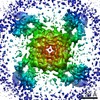 22661MC M: map data used to model this data C: citing same article ( |
|---|---|
| Similar structure data |
-Links
 PDBj
PDBj


- Assembly
| Deposited unit | 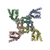
|
|---|---|
| 1 |
|
-Components
| #1: Protein | Mass: 68519.062 Da / Num. of mol.: 4 / Mutation: R398A,L506A,M513V Source method: isolated from a genetically manipulated source Source: (gene. exp.) Arcobacter butzleri (strain RM4018) (bacteria), (gene. exp.) Homo sapiens (human)Strain: K12, RM4018 / Gene: malE, b4034, JW3994, Abu_1752, SCN9A, NENA / Production host: Trichoplusia ni (cabbage looper)References: UniProt: P0AEX9, UniProt: A8EVM5, UniProt: Q15858 #2: Protein/peptide | Mass: 3999.777 Da / Num. of mol.: 4 / Mutation: E1G, E4G, Y33W / Source method: obtained synthetically / Source: (synth.) Haplopelma schmidti (Chinese earth tiger) / References: UniProt: P83303Has protein modification | Y | |
|---|
-Experimental details
-Experiment
| Experiment | Method: ELECTRON MICROSCOPY |
|---|---|
| EM experiment | Aggregation state: PARTICLE / 3D reconstruction method: single particle reconstruction |
- Sample preparation
| Component |
| |||||||||||||||||||||||||
|---|---|---|---|---|---|---|---|---|---|---|---|---|---|---|---|---|---|---|---|---|---|---|---|---|---|---|
| Molecular weight | Value: 0.3 MDa / Experimental value: NO | |||||||||||||||||||||||||
| Source (natural) |
| |||||||||||||||||||||||||
| Source (recombinant) |
| |||||||||||||||||||||||||
| Buffer solution | pH: 7.5 | |||||||||||||||||||||||||
| Buffer component |
| |||||||||||||||||||||||||
| Specimen | Conc.: 10 mg/ml / Embedding applied: NO / Shadowing applied: NO / Staining applied: NO / Vitrification applied: YES Details: m3-HwTx-IV was added in excess to the chimera at 8:1 stoichiometric molar ratio of toxin to channel. | |||||||||||||||||||||||||
| Specimen support | Grid material: GOLD / Grid mesh size: 400 divisions/in. / Grid type: Quantifoil R1.2/1.3 | |||||||||||||||||||||||||
| Vitrification | Instrument: FEI VITROBOT MARK IV / Cryogen name: ETHANE / Humidity: 100 % / Chamber temperature: 293 K / Details: Blot for 2.5-4.0 seconds before plunging |
- Electron microscopy imaging
| Experimental equipment |  Model: Titan Krios / Image courtesy: FEI Company |
|---|---|
| Microscopy | Model: FEI TITAN KRIOS / Details: Preliminary grid screening was performed manually. |
| Electron gun | Electron source:  FIELD EMISSION GUN / Accelerating voltage: 300 kV / Illumination mode: FLOOD BEAM FIELD EMISSION GUN / Accelerating voltage: 300 kV / Illumination mode: FLOOD BEAM |
| Electron lens | Mode: BRIGHT FIELD / Nominal magnification: 130000 X / Nominal defocus max: 2500 nm / Nominal defocus min: 500 nm / Cs: 2.7 mm / C2 aperture diameter: 100 µm / Alignment procedure: BASIC |
| Specimen holder | Cryogen: NITROGEN / Specimen holder model: FEI TITAN KRIOS AUTOGRID HOLDER / Temperature (max): 70 K / Temperature (min): 70 K |
| Image recording | Average exposure time: 8.6 sec. / Electron dose: 60 e/Å2 / Detector mode: SUPER-RESOLUTION / Film or detector model: GATAN K2 SUMMIT (4k x 4k) / Num. of real images: 7168 |
| EM imaging optics | Energyfilter name: GIF Bioquantum / Energyfilter slit width: 20 eV / Phase plate: VOLTA PHASE PLATE |
| Image scans | Width: 3710 / Height: 3582 / Movie frames/image: 42 / Used frames/image: 2-42 |
- Processing
| EM software |
| ||||||||||||||||||||||||||||||||||||
|---|---|---|---|---|---|---|---|---|---|---|---|---|---|---|---|---|---|---|---|---|---|---|---|---|---|---|---|---|---|---|---|---|---|---|---|---|---|
| CTF correction | Type: PHASE FLIPPING AND AMPLITUDE CORRECTION | ||||||||||||||||||||||||||||||||||||
| Particle selection | Num. of particles selected: 1470000 | ||||||||||||||||||||||||||||||||||||
| Symmetry | Point symmetry: C4 (4 fold cyclic) | ||||||||||||||||||||||||||||||||||||
| 3D reconstruction | Resolution: 3.6 Å / Resolution method: FSC 0.143 CUT-OFF / Num. of particles: 501310 / Algorithm: FOURIER SPACE / Num. of class averages: 1 / Symmetry type: POINT | ||||||||||||||||||||||||||||||||||||
| Atomic model building | Protocol: FLEXIBLE FIT / Space: REAL / Target criteria: Correlation coefficient | ||||||||||||||||||||||||||||||||||||
| Atomic model building | PDB-ID: 6N4I Pdb chain-ID: A / Accession code: 6N4I / Source name: PDB / Type: experimental model |