 Movie
Movie Controller

[English] 日本語
 Yorodumi
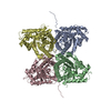
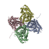
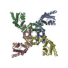
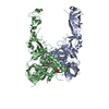
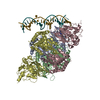
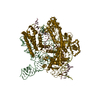
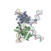
- PDB-6dzl: Ebola virus Makona variant GP (mucin-deleted) in complex with pan... -
+ Open data

- Basic information
| Entry | Database: PDB / ID: 6dzl | |||||||||
|---|---|---|---|---|---|---|---|---|---|---|
| Title | Ebola virus Makona variant GP (mucin-deleted) in complex with pan-ebolavirus human antibody ADI-15878 Fab | |||||||||
 Components Components |
| |||||||||
 Keywords Keywords | VIRAL PROTEIN / Ebola / antibody / pan-ebolavirus / internal fusion loop / HR1 / glycoprotein | |||||||||
| Function / homology |  Function and homology information Function and homology informationsymbiont-mediated-mediated suppression of host tetherin activity / clathrin-dependent endocytosis of virus by host cell / entry receptor-mediated virion attachment to host cell / symbiont-mediated suppression of host innate immune response / fusion of virus membrane with host endosome membrane / viral envelope / symbiont entry into host cell / lipid binding / host cell plasma membrane / virion membrane / extracellular region Similarity search - Function | |||||||||
| Biological species |   Zaire ebolavirus Zaire ebolavirus Homo sapiens (human) Homo sapiens (human) | |||||||||
| Method | ELECTRON MICROSCOPY / single particle reconstruction / cryo EM / Resolution: 4.14 Å | |||||||||
 Authors Authors | Murin, C.D. / Ward, A.B. | |||||||||
| Funding support |  United States, 1items United States, 1items
| |||||||||
 Citation Citation | Journal: Cell Rep / Year: 2018 Title: Structural Basis of Pan-Ebolavirus Neutralization by an Antibody Targeting the Glycoprotein Fusion Loop. Authors: Charles D Murin / Jessica F Bruhn / Zachary A Bornholdt / Jeffrey Copps / Robyn Stanfield / Andrew B Ward / Abstract: Monoclonal antibodies (mAbs) with pan-ebolavirus cross-reactivity are highly desirable, but development of such mAbs is limited by a lack of a molecular understanding of cross-reactive epitopes. The ...Monoclonal antibodies (mAbs) with pan-ebolavirus cross-reactivity are highly desirable, but development of such mAbs is limited by a lack of a molecular understanding of cross-reactive epitopes. The antibody ADI-15878 was previously identified from a human survivor of Ebola virus Makona variant (EBOV/Mak) infection. This mAb demonstrated potent neutralizing activity against all known ebolaviruses and provided protection in rodent and ferret models against three ebolavirus species. Here, we describe the unliganded crystal structure of ADI-15878 as well as the cryo-EM structures of ADI-15878 in complex with the EBOV/Mak and Bundibugyo virus (BDBV) glycoproteins (GPs). ADI-15878 binds through an induced-fit mechanism by targeting highly conserved residues in the internal fusion loop (IFL), bridging across GP protomers via the heptad repeat 1 (HR1) region. Our structures provide a more complete description of the ebolavirus immunogenic landscape, as well as a molecular basis for how rare but potent antibodies target conserved filoviral fusion machinery. | |||||||||
| History |
|
- Structure visualization
| Movie |
Movie viewer |
|---|---|
| Structure viewer | Molecule: MolmilJmol/JSmol |
- Downloads & links
-Download
| PDBx/mmCIF format | 6dzl.cif.gz | 295.5 KB | Display | PDBx/mmCIF format |
|---|---|---|---|---|
| PDB format | pdb6dzl.ent.gz | 236.2 KB | Display | PDB format |
| PDBx/mmJSON format | 6dzl.json.gz | Tree view | PDBx/mmJSON format | |
| Others |  Other downloads Other downloads |
-Validation report
| Arichive directory | https://data.pdbj.org/pub/pdb/validation_reports/dz/6dzlftp://data.pdbj.org/pub/pdb/validation_reports/dz/6dzl | HTTPS FTP |
|---|
-Related structure data
| Related structure data |  8935MC  8936C  6dzmC  6dznC M: map data used to model this data C: citing same article ( |
|---|---|
| Similar structure data |

-Links
 PDBj
PDBj


- Assembly
| Deposited unit | 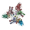
|
|---|---|
| 1 |
|
-Components
-Ebola virus Makona ... , 2 types, 6 molecules ABCDEF
| #1: Protein | Mass: 35517.750 Da / Num. of mol.: 3 Source method: isolated from a genetically manipulated source Source: (gene. exp.) Zaire ebolavirus / Strain: Kikwit-95 / Gene: GP / Variant: Makona / Plasmid: pPPI4 / Cell line (production host): HEK 293F / Production host: Homo sapiens (human) / References: UniProt: A0A0F7IM40, UniProt: P87666*PLUS#2: Protein | Mass: 18334.406 Da / Num. of mol.: 3 Source method: isolated from a genetically manipulated source Source: (gene. exp.) Zaire ebolavirus / Strain: Kikwit-95 / Gene: GP / Variant: Makona / Plasmid: pPPI4 / Cell line (production host): HEK 293F / Production host: Homo sapiens (human) / References: UniProt: P87666 |
|---|
-Antibody , 2 types, 6 molecules JKLGHI
| #3: Antibody | Mass: 11333.459 Da / Num. of mol.: 3 Source method: isolated from a genetically manipulated source Source: (gene. exp.) Homo sapiens (human) / Production host:  #4: Antibody | Mass: 13171.703 Da / Num. of mol.: 3 Source method: isolated from a genetically manipulated source Source: (gene. exp.) Homo sapiens (human) / Production host: |
|---|
-Sugars , 3 types, 12 molecules 
| #5: Polysaccharide | Source method: isolated from a genetically manipulated source #6: Polysaccharide | Source method: isolated from a genetically manipulated source #7: Sugar | ChemComp-NAG /  Type: D-saccharide, beta linking / Mass: 221.208 Da / Num. of mol.: 6 Type: D-saccharide, beta linking / Mass: 221.208 Da / Num. of mol.: 6Source method: isolated from a genetically manipulated source Formula: C8H15NO6 |
|---|
-Details
| Has protein modification | Y |
|---|
-Experimental details
-Experiment
| Experiment | Method: ELECTRON MICROSCOPY |
|---|---|
| EM experiment | Aggregation state: PARTICLE / 3D reconstruction method: single particle reconstruction |
- Sample preparation
| Component |
| ||||||||||||||||||||||||||||
|---|---|---|---|---|---|---|---|---|---|---|---|---|---|---|---|---|---|---|---|---|---|---|---|---|---|---|---|---|---|
| Molecular weight |
| ||||||||||||||||||||||||||||
| Source (natural) |
| ||||||||||||||||||||||||||||
| Source (recombinant) |
| ||||||||||||||||||||||||||||
| Buffer solution | pH: 7.4 Details: Solutions were made fresh from 10X concentrate and filtered with a 0.2 micrometer filter into a sterile glass container. | ||||||||||||||||||||||||||||
| Buffer component |
| ||||||||||||||||||||||||||||
| Specimen | Conc.: 1 mg/ml / Embedding applied: NO / Shadowing applied: NO / Staining applied: NO / Vitrification applied: YES Details: Antibody ADI-15878 Fab was added in 10 molar excess to glycoprotein and allowed to incubate overnight at 4 degrees Celsius followed by size exclusion chromatography to purify the complex and rid of excess Fab. | ||||||||||||||||||||||||||||
| Specimen support | Grid material: COPPER / Grid type: C-flat-1.2/1.3 4C | ||||||||||||||||||||||||||||
| Vitrification | Instrument: FEI VITROBOT MARK IV / Cryogen name: ETHANE / Humidity: 100 % / Chamber temperature: 277.15 K Details: 3.5s blot on both sides of the grid using Whattman No. 1 filter paper |
- Electron microscopy imaging
| Experimental equipment |  Model: Talos Arctica / Image courtesy: FEI Company |
|---|---|
| Microscopy | Model: FEI TALOS ARCTICA / Details: Preliminary grid screening was performed manually |
| Electron gun | Electron source:  FIELD EMISSION GUN / Accelerating voltage: 200 kV / Illumination mode: FLOOD BEAM FIELD EMISSION GUN / Accelerating voltage: 200 kV / Illumination mode: FLOOD BEAM |
| Electron lens | Mode: BRIGHT FIELD / Calibrated magnification: 43478 X / Nominal defocus max: 3500 nm / Nominal defocus min: 1000 nm / Cs: 2.7 mm / C2 aperture diameter: 100 µm / Alignment procedure: COMA FREE |
| Specimen holder | Cryogen: NITROGEN / Specimen holder model: FEI TITAN KRIOS AUTOGRID HOLDER |
| Image recording | Electron dose: 60 e/Å2 / Detector mode: COUNTING / Film or detector model: GATAN K2 SUMMIT (4k x 4k) |
- Processing
| Software | Name: PHENIX / Version: 1.13_2998: / Classification: refinement | ||||||||||||||||||||||||||||
|---|---|---|---|---|---|---|---|---|---|---|---|---|---|---|---|---|---|---|---|---|---|---|---|---|---|---|---|---|---|
| EM software |
| ||||||||||||||||||||||||||||
| CTF correction | Type: PHASE FLIPPING AND AMPLITUDE CORRECTION | ||||||||||||||||||||||||||||
| Symmetry | Point symmetry: C3 (3 fold cyclic) | ||||||||||||||||||||||||||||
| 3D reconstruction | Resolution: 4.14 Å / Resolution method: FSC 0.143 CUT-OFF / Num. of particles: 157861 / Symmetry type: POINT |
