 Movie
Movie Controller

[English] 日本語
 Yorodumi
- PDB-7e2w: Crystal structure of isocitrate dehydrogenase from Ostreococcus t... -
+ Open data

- Basic information
| Entry | Database: PDB / ID: 7e2w | |||||||||
|---|---|---|---|---|---|---|---|---|---|---|
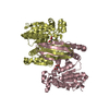
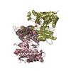
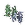
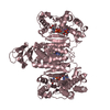
| Title | Crystal structure of isocitrate dehydrogenase from Ostreococcus tauri in complex with isocitrate and magnesium(II) | |||||||||
 Components Components | Isocitrate dehydrogenase (NAD(+)), mitochondrial | |||||||||
 Keywords Keywords | OXIDOREDUCTASE / isocitrate dehydrogenase | |||||||||
| Function / homology |  Function and homology information Function and homology informationisocitrate dehydrogenase (NAD+) / isocitrate dehydrogenase (NAD+) activity / isocitrate metabolic process / isocitrate dehydrogenase (NADP+) activity / NADP+ metabolic process / NAD+ metabolic process / tricarboxylic acid cycle / magnesium ion binding / protein homodimerization activity / mitochondrion Similarity search - Function | |||||||||
| Biological species |  Ostreococcus tauri (plant) Ostreococcus tauri (plant) | |||||||||
| Method |  X-RAY DIFFRACTION / SYNCHROTRON / MOLECULAR REPLACEMENT / molecular replacement / Resolution: 1.8 Å X-RAY DIFFRACTION / SYNCHROTRON / MOLECULAR REPLACEMENT / molecular replacement / Resolution: 1.8 Å | |||||||||
 Authors Authors | Zhu, G.P. / Tang, W.G. / Wang, P. | |||||||||
| Funding support |  China, 1items China, 1items
| |||||||||
 Citation Citation | Journal: Arch.Biochem.Biophys. / Year: 2021 Title: Crystal structures of NAD + -linked isocitrate dehydrogenase from the green alga Ostreococcus tauri and its evolutionary relationship with eukaryotic NADP + -linked homologs. Authors: Tang, W. / Wu, M. / Qin, N. / Liu, L. / Meng, R. / Wang, C. / Wang, P. / Zang, J. / Zhu, G. | |||||||||
| History |
|
- Structure visualization
| Structure viewer | Molecule: MolmilJmol/JSmol |
|---|
- Downloads & links
-Download
| PDBx/mmCIF format | 7e2w.cif.gz | 329.8 KB | Display | PDBx/mmCIF format |
|---|---|---|---|---|
| PDB format | pdb7e2w.ent.gz | 263.1 KB | Display | PDB format |
| PDBx/mmJSON format | 7e2w.json.gz | Tree view | PDBx/mmJSON format | |
| Others |  Other downloads Other downloads |
-Validation report
| Arichive directory | https://data.pdbj.org/pub/pdb/validation_reports/e2/7e2wftp://data.pdbj.org/pub/pdb/validation_reports/e2/7e2w | HTTPS FTP |
|---|
-Related structure data
| Related structure data |  6ixlSC 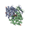 6ixnC  6ixtC S: Starting model for refinement C: citing same article ( |
|---|---|
| Similar structure data |


-Links
 PDBj
PDBj


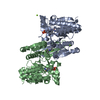
- Assembly
| Deposited unit | 
| ||||||||
|---|---|---|---|---|---|---|---|---|---|
| 1 | 
| ||||||||
| 2 | 
| ||||||||
| Unit cell |
|
-Components
-Protein , 1 types, 4 molecules ABCD
| #1: Protein | Mass: 46823.742 Da / Num. of mol.: 4 Source method: isolated from a genetically manipulated source Source: (gene. exp.) Ostreococcus tauri (plant) / Gene: IDH, Ot_13g02940, BE221DRAFT_192402 / Production host:  References: UniProt: A0A096P8D3, isocitrate dehydrogenase (NAD+) |
|---|
-Non-polymers , 6 types, 702 molecules 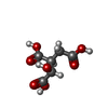


| #2: Chemical | ChemComp-ICT /  Mass: 192.124 Da / Num. of mol.: 1 / Source method: obtained synthetically / Formula: C6H8O7 / Feature type: SUBJECT OF INVESTIGATION Mass: 192.124 Da / Num. of mol.: 1 / Source method: obtained synthetically / Formula: C6H8O7 / Feature type: SUBJECT OF INVESTIGATION | ||||
|---|---|---|---|---|---|
| #3: Chemical | ChemComp-MG /  Mass: 24.305 Da / Num. of mol.: 1 / Source method: obtained synthetically / Formula: Mg / Feature type: SUBJECT OF INVESTIGATION Mass: 24.305 Da / Num. of mol.: 1 / Source method: obtained synthetically / Formula: Mg / Feature type: SUBJECT OF INVESTIGATION | ||||
| #4: Chemical | ChemComp-FLC /  Mass: 189.100 Da / Num. of mol.: 1 / Source method: obtained synthetically / Formula: C6H5O7 Mass: 189.100 Da / Num. of mol.: 1 / Source method: obtained synthetically / Formula: C6H5O7 | ||||
| #5: Chemical |  Mass: 60.095 Da / Num. of mol.: 3 / Source method: obtained synthetically / Formula: C3H8O Mass: 60.095 Da / Num. of mol.: 3 / Source method: obtained synthetically / Formula: C3H8O#6: Chemical |  Mass: 92.094 Da / Num. of mol.: 3 / Source method: isolated from a natural source / Formula: C3H8O3 Mass: 92.094 Da / Num. of mol.: 3 / Source method: isolated from a natural source / Formula: C3H8O3#7: Water | ChemComp-HOH / | Mass: 18.015 Da / Num. of mol.: 693 / Source method: isolated from a natural source / Formula: H2O |
-Details
| Has ligand of interest | Y |
|---|
-Experimental details
-Experiment
| Experiment | Method: X-RAY DIFFRACTION / Number of used crystals: 1 |
|---|
- Sample preparation
| Crystal | Density Matthews: 2.77 Å3/Da / Density % sol: 55.64 % |
|---|---|
| Crystal grow | Temperature: 286 K / Method: vapor diffusion, sitting drop / pH: 5.5 Details: 20% PEG 4000, 10% 2-propanol, 0.1 M sodium citrate (pH 5.5). |
-Data collection
| Diffraction | Mean temperature: 100 K / Serial crystal experiment: N |
|---|---|
| Diffraction source | Source: SYNCHROTRON / Site: SSRF / Beamline: BL17U1 / Wavelength: 0.97915 Å |
| Detector | Type: ADSC QUANTUM 315r / Detector: CCD / Date: Apr 25, 2015 |
| Radiation | Protocol: SINGLE WAVELENGTH / Monochromatic (M) / Laue (L): M / Scattering type: x-ray |
| Radiation wavelength | Wavelength: 0.97915 Å / Relative weight: 1 |
| Reflection | Resolution: 1.8→20 Å / Num. obs: 177015 / % possible obs: 94.9 % / Redundancy: 2.2 % / CC1/2: 0.998 / Rmerge(I) obs: 0.039 / Net I/σ(I): 14.4 |
| Reflection shell | Resolution: 1.8→1.9 Å / Num. unique obs: 26019 / CC1/2: 0.84 |
-Phasing
| Phasing | Method: molecular replacement |
|---|
- Processing
| Software |
| ||||||||||||||||||||||||||||||||||||||||||||||||||||||||||||
|---|---|---|---|---|---|---|---|---|---|---|---|---|---|---|---|---|---|---|---|---|---|---|---|---|---|---|---|---|---|---|---|---|---|---|---|---|---|---|---|---|---|---|---|---|---|---|---|---|---|---|---|---|---|---|---|---|---|---|---|---|---|
| Refinement | Method to determine structure: MOLECULAR REPLACEMENT Starting model: 6IXL Resolution: 1.8→19.95 Å / Cor.coef. Fo:Fc: 0.951 / Cor.coef. Fo:Fc free: 0.937 / SU B: 3.355 / SU ML: 0.1 / Cross valid method: THROUGHOUT / σ(F): 0 / ESU R: 0.13 / ESU R Free: 0.124 / Stereochemistry target values: MAXIMUM LIKELIHOOD Details: HYDROGENS HAVE BEEN ADDED IN THE RIDING POSITIONS U VALUES : REFINED INDIVIDUALLY
| ||||||||||||||||||||||||||||||||||||||||||||||||||||||||||||
| Solvent computation | Ion probe radii: 0.8 Å / Shrinkage radii: 0.8 Å / VDW probe radii: 1.2 Å / Solvent model: MASK | ||||||||||||||||||||||||||||||||||||||||||||||||||||||||||||
| Displacement parameters | Biso max: 94.09 Å2 / Biso mean: 29.875 Å2 / Biso min: 14.8 Å2
| ||||||||||||||||||||||||||||||||||||||||||||||||||||||||||||
| Refinement step | Cycle: final / Resolution: 1.8→19.95 Å
| ||||||||||||||||||||||||||||||||||||||||||||||||||||||||||||
| Refine LS restraints |
| ||||||||||||||||||||||||||||||||||||||||||||||||||||||||||||
| LS refinement shell | Resolution: 1.8→1.847 Å / Rfactor Rfree error: 0 / Total num. of bins used: 20
|
