 Movie
Movie Controller


+ Open data

- Basic information
| Entry | Database: PDB / ID: 6adg | ||||||
|---|---|---|---|---|---|---|---|
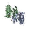
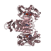
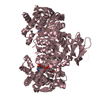
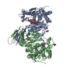
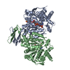
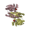
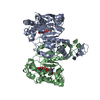
| Title | Crystal Structures of IDH1 R132H in complex with AG-881 | ||||||
 Components Components | Isocitrate dehydrogenase [NADP] cytoplasmic | ||||||
 Keywords Keywords | OXIDOREDUCTASE / IDH1 | ||||||
| Function / homology |  Function and homology information Function and homology informationAbnormal conversion of 2-oxoglutarate to 2-hydroxyglutarate / NADPH regeneration / NFE2L2 regulating TCA cycle genes / isocitrate metabolic process / isocitrate dehydrogenase (NADP+) / NADPH regeneration / isocitrate dehydrogenase (NADP+) activity / NADP+ metabolic process / 2-oxoglutarate metabolic process / glyoxylate cycle ...Abnormal conversion of 2-oxoglutarate to 2-hydroxyglutarate / NADPH regeneration / NFE2L2 regulating TCA cycle genes / isocitrate metabolic process / isocitrate dehydrogenase (NADP+) / NADPH regeneration / isocitrate dehydrogenase (NADP+) activity / NADP+ metabolic process / 2-oxoglutarate metabolic process / glyoxylate cycle / response to steroid hormone / female gonad development / peroxisomal matrix / tricarboxylic acid cycle / Peroxisomal protein import / NAD binding / NADP binding / tertiary granule lumen / peroxisome / secretory granule lumen / ficolin-1-rich granule lumen / cadherin binding / Neutrophil degranulation / magnesium ion binding / protein homodimerization activity / mitochondrion / extracellular exosome / extracellular region / identical protein binding / cytosol / cytoplasm Similarity search - Function | ||||||
| Biological species |  Homo sapiens (human) Homo sapiens (human) | ||||||
| Method |  X-RAY DIFFRACTION / SYNCHROTRON / MOLECULAR REPLACEMENT / Resolution: 3 Å X-RAY DIFFRACTION / SYNCHROTRON / MOLECULAR REPLACEMENT / Resolution: 3 Å | ||||||
 Authors Authors | Ma, R. / Yun, C.H. | ||||||
 Citation Citation | Journal: Biochem. Biophys. Res. Commun. / Year: 2018 Title: Crystal structures of pan-IDH inhibitor AG-881 in complex with mutant human IDH1 and IDH2 Authors: Ma, R. / Yun, C.H. | ||||||
| History |
|
- Structure visualization
| Structure viewer | Molecule: MolmilJmol/JSmol |
|---|
- Downloads & links
-Download
| PDBx/mmCIF format | 6adg.cif.gz | 248.8 KB | Display | PDBx/mmCIF format |
|---|---|---|---|---|
| PDB format | pdb6adg.ent.gz | 198.8 KB | Display | PDB format |
| PDBx/mmJSON format | 6adg.json.gz | Tree view | PDBx/mmJSON format | |
| Others |  Other downloads Other downloads |
-Validation report
| Arichive directory | https://data.pdbj.org/pub/pdb/validation_reports/ad/6adgftp://data.pdbj.org/pub/pdb/validation_reports/ad/6adg | HTTPS FTP |
|---|
-Related structure data
| Related structure data |  6adiC  5de1S S: Starting model for refinement C: citing same article ( |
|---|---|
| Similar structure data |


-Links
 PDBj
PDBj


- Assembly
| Deposited unit | 
| ||||||||
|---|---|---|---|---|---|---|---|---|---|
| 1 | 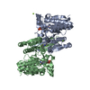
| ||||||||
| 2 | 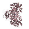
| ||||||||
| Unit cell |
| ||||||||
| Components on special symmetry positions |
|
-Components
| #1: Protein | Mass: 48135.684 Da / Num. of mol.: 3 / Mutation: R132H Source method: isolated from a genetically manipulated source Source: (gene. exp.) Homo sapiens (human) / Gene: IDH1 / Production host:   Spodoptera frugiperda (fall armyworm) Spodoptera frugiperda (fall armyworm)References: UniProt: O75874, isocitrate dehydrogenase (NADP+) #2: Chemical |   Mass: 745.421 Da / Num. of mol.: 2 Mass: 745.421 Da / Num. of mol.: 2Source method: isolated from a genetically manipulated source Formula: C21H30N7O17P3 #3: Chemical |   Mass: 24.305 Da / Num. of mol.: 2 Mass: 24.305 Da / Num. of mol.: 2Source method: isolated from a genetically manipulated source Formula: Mg #4: Chemical |   Mass: 414.737 Da / Num. of mol.: 2 / Source method: obtained synthetically / Formula: C14H13ClF6N6 Mass: 414.737 Da / Num. of mol.: 2 / Source method: obtained synthetically / Formula: C14H13ClF6N6#5: Water | ChemComp-HOH / |  Mass: 18.015 Da / Num. of mol.: 121 / Source method: isolated from a natural source / Formula: H2O Mass: 18.015 Da / Num. of mol.: 121 / Source method: isolated from a natural source / Formula: H2O |
|---|
-Experimental details
-Experiment
| Experiment | Method: X-RAY DIFFRACTION / Number of used crystals: 1 |
|---|
- Sample preparation
| Crystal | Density Matthews: 2.69 Å3/Da / Density % sol: 54.31 % |
|---|---|
| Crystal grow | Temperature: 293 K / Method: vapor diffusion, hanging drop / pH: 8.5 Details: 0.2M Magnesium chloride hexahydrate, 0.1M Tris pH 8.5, 25%(w/v) Polyethylene glycol 3,350, 0.1M Cesium chloride |
-Data collection
| Diffraction | Mean temperature: 100 K |
|---|---|
| Diffraction source | Source: SYNCHROTRON / Site: SSRF  / Beamline: BL17U1 / Wavelength: 0.97918 Å / Beamline: BL17U1 / Wavelength: 0.97918 Å |
| Detector | Type: DECTRIS EIGER X 16M / Detector: PIXEL / Date: Nov 30, 2017 |
| Radiation | Protocol: SINGLE WAVELENGTH / Monochromatic (M) / Laue (L): M / Scattering type: x-ray |
| Radiation wavelength | Wavelength: 0.97918 Å / Relative weight: 1 |
| Reflection | Resolution: 3→50 Å / Num. obs: 30674 / % possible obs: 92.6 % / Redundancy: 3.8 % / Rpim(I) all: 0.105 / Net I/σ(I): 5.4 |
| Reflection shell | Resolution: 3→3.23 Å / Num. unique obs: 5478 / Rpim(I) all: 0.436 |
- Processing
| Software |
| ||||||||||||||||||||||||||||||||||||||||||||||||||||||||||||||||||||||||||||||||||||
|---|---|---|---|---|---|---|---|---|---|---|---|---|---|---|---|---|---|---|---|---|---|---|---|---|---|---|---|---|---|---|---|---|---|---|---|---|---|---|---|---|---|---|---|---|---|---|---|---|---|---|---|---|---|---|---|---|---|---|---|---|---|---|---|---|---|---|---|---|---|---|---|---|---|---|---|---|---|---|---|---|---|---|---|---|---|
| Refinement | Method to determine structure: MOLECULAR REPLACEMENT Starting model: 5DE1 Resolution: 3→43.015 Å / SU ML: 0.39 / Cross valid method: FREE R-VALUE / σ(F): 0.31 / Phase error: 24.1
| ||||||||||||||||||||||||||||||||||||||||||||||||||||||||||||||||||||||||||||||||||||
| Solvent computation | Shrinkage radii: 0.9 Å / VDW probe radii: 1.11 Å | ||||||||||||||||||||||||||||||||||||||||||||||||||||||||||||||||||||||||||||||||||||
| Refinement step | Cycle: LAST / Resolution: 3→43.015 Å
| ||||||||||||||||||||||||||||||||||||||||||||||||||||||||||||||||||||||||||||||||||||
| Refine LS restraints |
| ||||||||||||||||||||||||||||||||||||||||||||||||||||||||||||||||||||||||||||||||||||
| LS refinement shell |
|
