 Movie
Movie Controller

[English] 日本語
 Yorodumi
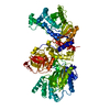
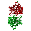
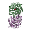
- PDB-6zyh: Crystal structure of GRP78 (70kDa heat shock protein 5 / BiP) ATP... -
+ Open data

- Basic information
| Entry | Database: PDB / ID: 6zyh | ||||||
|---|---|---|---|---|---|---|---|
| Title | Crystal structure of GRP78 (70kDa heat shock protein 5 / BiP) ATPase domain in complex with ADP and calcium | ||||||
 Components Components | Endoplasmic reticulum chaperone BiP | ||||||
 Keywords Keywords | CHAPERONE / BiP / HSP70 / GRP78 / ATPase domain / NBD / ADP | ||||||
| Function / homology |  Function and homology information Function and homology informationnegative regulation of IRE1-mediated unfolded protein response / post-translational protein targeting to membrane, translocation / non-chaperonin molecular chaperone ATPase / negative regulation of protein-containing complex assembly / ATP-dependent protein folding chaperone / melanosome / endoplasmic reticulum lumen / negative regulation of apoptotic process / endoplasmic reticulum membrane / perinuclear region of cytoplasm ...negative regulation of IRE1-mediated unfolded protein response / post-translational protein targeting to membrane, translocation / non-chaperonin molecular chaperone ATPase / negative regulation of protein-containing complex assembly / ATP-dependent protein folding chaperone / melanosome / endoplasmic reticulum lumen / negative regulation of apoptotic process / endoplasmic reticulum membrane / perinuclear region of cytoplasm / cell surface / ATP hydrolysis activity / mitochondrion / ATP binding / cytosol Similarity search - Function | ||||||
| Biological species |   Cricetulus griseus (Chinese hamster) Cricetulus griseus (Chinese hamster) | ||||||
| Method |  X-RAY DIFFRACTION / SYNCHROTRON / MOLECULAR REPLACEMENT / Resolution: 1.88 Å X-RAY DIFFRACTION / SYNCHROTRON / MOLECULAR REPLACEMENT / Resolution: 1.88 Å | ||||||
 Authors Authors | Yan, Y. / Preissler, S. / Ron, D. | ||||||
| Funding support |  United Kingdom, 1items United Kingdom, 1items
| ||||||
 Citation Citation | Journal: Elife / Year: 2020 Title: Calcium depletion challenges endoplasmic reticulum proteostasis by destabilising BiP-substrate complexes. Authors: Preissler, S. / Rato, C. / Yan, Y. / Perera, L.A. / Czako, A. / Ron, D. | ||||||
| History |
|
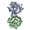
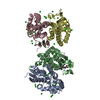
- Structure visualization
| Structure viewer | Molecule: MolmilJmol/JSmol |
|---|
- Downloads & links
-Download
| PDBx/mmCIF format | 6zyh.cif.gz | 165.1 KB | Display | PDBx/mmCIF format |
|---|---|---|---|---|
| PDB format | pdb6zyh.ent.gz | 127 KB | Display | PDB format |
| PDBx/mmJSON format | 6zyh.json.gz | Tree view | PDBx/mmJSON format | |
| Others |  Other downloads Other downloads |
-Validation report
| Arichive directory | https://data.pdbj.org/pub/pdb/validation_reports/zy/6zyhftp://data.pdbj.org/pub/pdb/validation_reports/zy/6zyh | HTTPS FTP |
|---|
-Related structure data
| Related structure data | 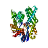 6zyiC 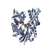 6zyjC 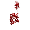 7a4uC 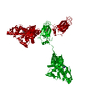 7a4vC  3iucS S: Starting model for refinement C: citing same article ( |
|---|---|
| Similar structure data |


-Links
 PDBj
PDBj


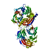
- Assembly
| Deposited unit | 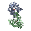
| ||||||||
|---|---|---|---|---|---|---|---|---|---|
| 1 |
| ||||||||
| Unit cell |
|
-Components
| #1: Protein | Mass: 41868.445 Da / Num. of mol.: 2 Source method: isolated from a genetically manipulated source Source: (gene. exp.) Cricetulus griseus (Chinese hamster) / Gene: HSPA5, GRP78, I79_019946 / Production host:  References: UniProt: G3I8R9, non-chaperonin molecular chaperone ATPase #2: Chemical |   Mass: 427.201 Da / Num. of mol.: 2 / Source method: obtained synthetically / Formula: C10H15N5O10P2 / Feature type: SUBJECT OF INVESTIGATION / Comment: ADP, energy-carrying molecule*YM Mass: 427.201 Da / Num. of mol.: 2 / Source method: obtained synthetically / Formula: C10H15N5O10P2 / Feature type: SUBJECT OF INVESTIGATION / Comment: ADP, energy-carrying molecule*YM#3: Chemical | ChemComp-CA /   Mass: 40.078 Da / Num. of mol.: 4 / Source method: obtained synthetically / Formula: Ca / Feature type: SUBJECT OF INVESTIGATION Mass: 40.078 Da / Num. of mol.: 4 / Source method: obtained synthetically / Formula: Ca / Feature type: SUBJECT OF INVESTIGATION#4: Water | ChemComp-HOH / |  Mass: 18.015 Da / Num. of mol.: 218 / Source method: isolated from a natural source / Formula: H2O Mass: 18.015 Da / Num. of mol.: 218 / Source method: isolated from a natural source / Formula: H2OHas ligand of interest | Y | |
|---|
-Experimental details
-Experiment
| Experiment | Method: X-RAY DIFFRACTION / Number of used crystals: 1 |
|---|
- Sample preparation
| Crystal | Density Matthews: 2.37 Å3/Da / Density % sol: 48.06 % |
|---|---|
| Crystal grow | Temperature: 293 K / Method: vapor diffusion, sitting drop / pH: 5 / Details: 26% PEG6000, 0.2M CaCl2, 0.1MNaOAc Ph5 |
-Data collection
| Diffraction | Mean temperature: 100 K / Serial crystal experiment: N |
|---|---|
| Diffraction source | Source: SYNCHROTRON / Site: Diamond / Beamline: I03 / Wavelength: 0.97628 Å |
| Detector | Type: DECTRIS PILATUS 6M-F / Detector: PIXEL / Date: Jun 24, 2019 |
| Radiation | Protocol: SINGLE WAVELENGTH / Monochromatic (M) / Laue (L): M / Scattering type: x-ray |
| Radiation wavelength | Wavelength: 0.97628 Å / Relative weight: 1 |
| Reflection | Resolution: 1.88→29.14 Å / Num. obs: 62200 / % possible obs: 97.4 % / Redundancy: 8.9 % / CC1/2: 0.999 / Rmerge(I) obs: 0.063 / Net I/σ(I): 18.2 |
| Reflection shell | Resolution: 1.88→1.92 Å / Rmerge(I) obs: 0.989 / Num. unique obs: 3792 / CC1/2: 0.669 |
- Processing
| Software |
| ||||||||||||||||||||||||||||||||||||||||||||||||||||||||||||
|---|---|---|---|---|---|---|---|---|---|---|---|---|---|---|---|---|---|---|---|---|---|---|---|---|---|---|---|---|---|---|---|---|---|---|---|---|---|---|---|---|---|---|---|---|---|---|---|---|---|---|---|---|---|---|---|---|---|---|---|---|---|
| Refinement | Method to determine structure: MOLECULAR REPLACEMENT Starting model: 3IUC Resolution: 1.88→29.13 Å / Cor.coef. Fo:Fc: 0.954 / Cor.coef. Fo:Fc free: 0.948 / SU B: 3.557 / SU ML: 0.104 / Cross valid method: THROUGHOUT / σ(F): 0 / ESU R: 0.17 / ESU R Free: 0.14 / Stereochemistry target values: MAXIMUM LIKELIHOOD Details: HYDROGENS HAVE BEEN ADDED IN THE RIDING POSITIONS U VALUES : REFINED INDIVIDUALLY
| ||||||||||||||||||||||||||||||||||||||||||||||||||||||||||||
| Solvent computation | Ion probe radii: 0.8 Å / Shrinkage radii: 0.8 Å / VDW probe radii: 1.2 Å / Solvent model: MASK | ||||||||||||||||||||||||||||||||||||||||||||||||||||||||||||
| Displacement parameters | Biso max: 73.74 Å2 / Biso mean: 32.466 Å2 / Biso min: 21.28 Å2
| ||||||||||||||||||||||||||||||||||||||||||||||||||||||||||||
| Refinement step | Cycle: final / Resolution: 1.88→29.13 Å
| ||||||||||||||||||||||||||||||||||||||||||||||||||||||||||||
| Refine LS restraints |
| ||||||||||||||||||||||||||||||||||||||||||||||||||||||||||||
| LS refinement shell | Resolution: 1.88→1.929 Å / Rfactor Rfree error: 0 / Total num. of bins used: 20
|
