 Movie
Movie Controller


+ Open data

- Basic information
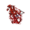
| Entry | Database: PDB / ID: 2dik | |||||||||
|---|---|---|---|---|---|---|---|---|---|---|
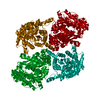
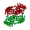
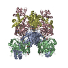
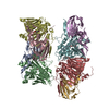
| Title | R337A MUTANT OF PYRUVATE PHOSPHATE DIKINASE | |||||||||
 Components Components | PROTEIN (PYRUVATE PHOSPHATE DIKINASE) | |||||||||
 Keywords Keywords | TRANSFERASE / PHOSPHOTRANSFERASE / KINASE | |||||||||
| Function / homology |  Function and homology information Function and homology informationpyruvate, phosphate dikinase / pyruvate, phosphate dikinase activity / kinase activity / ATP binding / metal ion binding Similarity search - Function | |||||||||
| Biological species |  Clostridium symbiosum (bacteria) Clostridium symbiosum (bacteria) | |||||||||
| Method |  X-RAY DIFFRACTION / MOLECULAR REPLACEMENT / Resolution: 2.5 Å X-RAY DIFFRACTION / MOLECULAR REPLACEMENT / Resolution: 2.5 Å | |||||||||
 Authors Authors | Huang, K. / Herzberg, O. | |||||||||
 Citation Citation | Journal: Biochemistry / Year: 1998 Title: Location of the phosphate binding site within Clostridium symbiosum pyruvate phosphate dikinase. Authors: McGuire, M. / Huang, K. / Kapadia, G. / Herzberg, O. / Dunaway-Mariano, D. #1: Journal: Proc.Natl.Acad.Sci.USA / Year: 1996Title: Swiveling-Domain Mechanism for Enzymatic Phosphotransfer between Remote Reaction Sites Authors: Herzberg, O. / Cheng, C.C.H. / Kapadia, G. / Mcguire, M. / Carroll, L.J. / Noh, S.J. / Dunaway-Mariano, D. | |||||||||
| History |
|
- Structure visualization
| Structure viewer | Molecule: MolmilJmol/JSmol |
|---|
- Downloads & links
-Download
| PDBx/mmCIF format | 2dik.cif.gz | 179.9 KB | Display | PDBx/mmCIF format |
|---|---|---|---|---|
| PDB format | pdb2dik.ent.gz | 141.3 KB | Display | PDB format |
| PDBx/mmJSON format | 2dik.json.gz | Tree view | PDBx/mmJSON format | |
| Others |  Other downloads Other downloads |
-Validation report
| Arichive directory | https://data.pdbj.org/pub/pdb/validation_reports/di/2dikftp://data.pdbj.org/pub/pdb/validation_reports/di/2dik | HTTPS FTP |
|---|
-Related structure data
| Related structure data |  1dikS S: Starting model for refinement |
|---|---|
| Similar structure data |


-Links
 PDBj
PDBj


- Assembly
| Deposited unit | 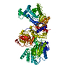
| ||||||||
|---|---|---|---|---|---|---|---|---|---|
| 1 | 
| ||||||||
| 2 | 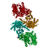
| ||||||||
| Unit cell |
| ||||||||
| Components on special symmetry positions |
|
-Components
| #1: Protein | Mass: 96568.055 Da / Num. of mol.: 1 / Mutation: R337A Source method: isolated from a genetically manipulated source Source: (gene. exp.) Clostridium symbiosum (bacteria) / Strain: JM 101 / Gene: PPDK / Plasmid: PACYC184D-12 / Gene (production host): PPDK / Production host: | ||
|---|---|---|---|
| #2: Chemical |   Mass: 96.063 Da / Num. of mol.: 2 / Source method: obtained synthetically / Formula: SO4 Mass: 96.063 Da / Num. of mol.: 2 / Source method: obtained synthetically / Formula: SO4#3: Water | ChemComp-HOH / |  Mass: 18.015 Da / Num. of mol.: 44 / Source method: isolated from a natural source / Formula: H2O Mass: 18.015 Da / Num. of mol.: 44 / Source method: isolated from a natural source / Formula: H2O |
-Experimental details
-Experiment
| Experiment | Method: X-RAY DIFFRACTION / Number of used crystals: 1 |
|---|
- Sample preparation
| Crystal | Density Matthews: 2.78 Å3/Da / Density % sol: 55.72 % | ||||||||||||||||||||||||||||||||||||||||||||||||
|---|---|---|---|---|---|---|---|---|---|---|---|---|---|---|---|---|---|---|---|---|---|---|---|---|---|---|---|---|---|---|---|---|---|---|---|---|---|---|---|---|---|---|---|---|---|---|---|---|---|
| Crystal grow | Temperature: 303 K / pH: 7 / Details: pH 7.0, temperature 303K | ||||||||||||||||||||||||||||||||||||||||||||||||
| Crystal grow | *PLUS Temperature: 30 ℃ / pH: 6.5 / Method: vapor diffusion, hanging drop | ||||||||||||||||||||||||||||||||||||||||||||||||
| Components of the solutions | *PLUS
|
-Data collection
| Diffraction | Mean temperature: 100 K |
|---|---|
| Diffraction source | Source: ROTATING ANODE / Type: RIGAKU RU200 / Wavelength: 1.5418 |
| Detector | Type: SIEMENS / Detector: AREA DETECTOR |
| Radiation | Monochromator: NI FILTER / Protocol: SINGLE WAVELENGTH / Monochromatic (M) / Laue (L): M / Scattering type: x-ray |
| Radiation wavelength | Wavelength: 1.5418 Å / Relative weight: 1 |
| Reflection | Resolution: 2.5→99 Å / Num. obs: 35455 / % possible obs: 95 % / Observed criterion σ(I): 1.2 / Redundancy: 2.8 % / Rsym value: 9.9 |
| Reflection shell | Resolution: 2.5→2.66 Å / % possible all: 83 |
| Reflection | *PLUS % possible obs: 95 % / Num. measured all: 97563 / Rmerge(I) obs: 0.099 |
| Reflection shell | *PLUS Highest resolution: 2.5 Å / % possible obs: 83 % / Mean I/σ(I) obs: 1.2 |
- Processing
| Software |
| ||||||||||||||||||||||||||||||
|---|---|---|---|---|---|---|---|---|---|---|---|---|---|---|---|---|---|---|---|---|---|---|---|---|---|---|---|---|---|---|---|
| Refinement | Method to determine structure: MOLECULAR REPLACEMENT Starting model: 1DIK Resolution: 2.5→8 Å / σ(F): 3
| ||||||||||||||||||||||||||||||
| Refinement step | Cycle: LAST / Resolution: 2.5→8 Å
| ||||||||||||||||||||||||||||||
| Refine LS restraints |
| ||||||||||||||||||||||||||||||
| Software | *PLUS Name: TNT / Classification: refinement | ||||||||||||||||||||||||||||||
| Refinement | *PLUS Highest resolution: 2.5 Å / σ(F): 3 / Rfactor obs: 0.18 | ||||||||||||||||||||||||||||||
| Solvent computation | *PLUS | ||||||||||||||||||||||||||||||
| Displacement parameters | *PLUS | ||||||||||||||||||||||||||||||
| Refine LS restraints | *PLUS Type: t_angle_deg / Dev ideal: 2.79 |
