 Movie
Movie Controller


+ Open data

- Basic information
| Entry | Database: PDB / ID: 1kbl | ||||||
|---|---|---|---|---|---|---|---|
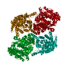
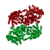
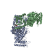
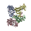
| Title | PYRUVATE PHOSPHATE DIKINASE | ||||||
 Components Components | PYRUVATE PHOSPHATE DIKINASE | ||||||
 Keywords Keywords | TRANSFERASE / PHOSPHOTRANSFERASE / KINASE | ||||||
| Function / homology |  Function and homology information Function and homology informationpyruvate, phosphate dikinase / pyruvate, phosphate dikinase activity / kinase activity / ATP binding / metal ion binding Similarity search - Function | ||||||
| Biological species |  Clostridium symbiosum (bacteria) Clostridium symbiosum (bacteria) | ||||||
| Method |  X-RAY DIFFRACTION / SYNCHROTRON / FOURIER SYNTHESIS / Resolution: 1.94 Å X-RAY DIFFRACTION / SYNCHROTRON / FOURIER SYNTHESIS / Resolution: 1.94 Å | ||||||
 Authors Authors | Herzberg, O. / Chen, C.C. / Liu, S. | ||||||
 Citation Citation | Journal: Biochemistry / Year: 2002 Title: Pyruvate site of pyruvate phosphate dikinase: crystal structure of the enzyme-phosphonopyruvate complex, and mutant analysis Authors: Herzberg, O. / Chen, C.C. / Liu, S. / Tempczyk, A. / Howard, A. / Wei, M. / Ye, D. / Dunaway-Mariano, D. #1: Journal: Proc.Natl.Acad.Sci.USA / Year: 1996Title: Swiveling-Domain Mechanism for Enzymatic Phosphotransfer between Remote Reaction Sites Authors: Herzberg, O. / Chen, C.C. / Kapadia, G. / Mcguire, M. / Carroll, L.J. / Noh, S.J. / Dunaway-Mariano, D. #2: Journal: Biochemistry / Year: 1998Title: Location of the Phosphate Binding Site within Clostridium Symbiosum Pyruvate Phosphate Dikinase Authors: Mcguire, M. / Huang, K. / Kapadia, G. / Herzberg, O. / Dunaway-Mariano, D. #3: Journal: J.Biol.Chem. / Year: 2000Title: Identification of Domain-Domain Docking Sites within Clostridium Symbiosum Pyruvate Phosphate Dikinase by Amino Acid Replacement Authors: Wei, M. / Li, Z. / Ye, D. / Herzberg, O. / Dunaway-Mariano, D. | ||||||
| History |
|
- Structure visualization
| Structure viewer | Molecule: MolmilJmol/JSmol |
|---|
- Downloads & links
-Download
| PDBx/mmCIF format | 1kbl.cif.gz | 203.1 KB | Display | PDBx/mmCIF format |
|---|---|---|---|---|
| PDB format | pdb1kbl.ent.gz | 158 KB | Display | PDB format |
| PDBx/mmJSON format | 1kbl.json.gz | Tree view | PDBx/mmJSON format | |
| Others |  Other downloads Other downloads |
-Validation report
| Arichive directory | https://data.pdbj.org/pub/pdb/validation_reports/kb/1kblftp://data.pdbj.org/pub/pdb/validation_reports/kb/1kbl | HTTPS FTP |
|---|
-Related structure data
| Related structure data |  1kc7SC S: Starting model for refinement C: citing same article ( |
|---|---|
| Similar structure data |


-Links
 PDBj
PDBj


- Assembly
| Deposited unit | 
| |||||||||
|---|---|---|---|---|---|---|---|---|---|---|
| 1 | 
| |||||||||
| 2 | 
| |||||||||
| Unit cell |
| |||||||||
| Components on special symmetry positions |
| |||||||||
| Details | A dimer generated by a crystallographic 2-fold axis: -1.0 0.0 0.0 89.837 0.0 1.0 0.0 0.0 0.0 0.0 -1.0 0.0 |
-Components
| #1: Protein | Mass: 96640.148 Da / Num. of mol.: 1 Source method: isolated from a genetically manipulated source Source: (gene. exp.) Clostridium symbiosum (bacteria) / Gene: PPDK / Plasmid: PACYC184-D12 / Gene (production host): PPDK / Production host: | ||
|---|---|---|---|
| #2: Chemical | ChemComp-NH4 /   Mass: 18.038 Da / Num. of mol.: 1 / Source method: obtained synthetically / Formula: H4N Mass: 18.038 Da / Num. of mol.: 1 / Source method: obtained synthetically / Formula: H4N | ||
| #3: Chemical | ChemComp-SO4 /   Mass: 96.063 Da / Num. of mol.: 5 / Source method: obtained synthetically / Formula: SO4 Mass: 96.063 Da / Num. of mol.: 5 / Source method: obtained synthetically / Formula: SO4#4: Water | ChemComp-HOH / |  Mass: 18.015 Da / Num. of mol.: 879 / Source method: isolated from a natural source / Formula: H2O Mass: 18.015 Da / Num. of mol.: 879 / Source method: isolated from a natural source / Formula: H2O |
-Experimental details
-Experiment
| Experiment | Method: X-RAY DIFFRACTION / Number of used crystals: 1 |
|---|
- Sample preparation
| Crystal | Density Matthews: 2.81 Å3/Da / Density % sol: 56.6 % | ||||||||||||||||||||||||||||||||||||||||||
|---|---|---|---|---|---|---|---|---|---|---|---|---|---|---|---|---|---|---|---|---|---|---|---|---|---|---|---|---|---|---|---|---|---|---|---|---|---|---|---|---|---|---|---|
| Crystal grow | Temperature: 303 K / Method: vapor diffusion, hanging drop / pH: 7 Details: 50-54% saturated ammonium sulfate, 10 mg/ml protein solution in 100mM KCl and 20mM imidazole buffer (pH 6.5), 100 mM Hepes buffer (pH 7.0), VAPOR DIFFUSION, HANGING DROP, temperature 303K | ||||||||||||||||||||||||||||||||||||||||||
| Crystal grow | *PLUS Temperature: 30 ℃Details: Herzberg, O., (1996) Proc.Natl.Acad.Sci.USA, 93, 2652. | ||||||||||||||||||||||||||||||||||||||||||
| Components of the solutions | *PLUS
|
-Data collection
| Diffraction | Mean temperature: 100 K |
|---|---|
| Diffraction source | Source: SYNCHROTRON / Site: NSLS  / Beamline: X12C / Wavelength: 1.3 / Wavelength: 1.3 Å / Beamline: X12C / Wavelength: 1.3 / Wavelength: 1.3 Å |
| Detector | Type: MARRESEARCH / Detector: IMAGE PLATE / Date: Jun 10, 1995 |
| Radiation | Monochromator: toroidal mirror / Protocol: SINGLE WAVELENGTH / Monochromatic (M) / Laue (L): M / Scattering type: x-ray |
| Radiation wavelength | Wavelength: 1.3 Å / Relative weight: 1 |
| Reflection | Resolution: 1.9→20 Å / Num. all: 79262 / Num. obs: 79262 / % possible obs: 93.5 % / Observed criterion σ(F): 0 / Observed criterion σ(I): 0 / Rmerge(I) obs: 0.051 / Rsym value: 0.051 / Net I/σ(I): 23.7 |
| Reflection shell | Resolution: 1.9→1.97 Å / Rmerge(I) obs: 0.405 / Mean I/σ(I) obs: 2.9 / Num. unique all: 6736 / % possible all: 80.1 |
| Reflection | *PLUS |
| Reflection shell | *PLUS % possible obs: 80.1 % / Num. unique obs: 6736 / Mean I/σ(I) obs: 2.9 |
- Processing
| Software |
| ||||||||||||||||||||||||||||||||||||||||||||||||||||||||||||
|---|---|---|---|---|---|---|---|---|---|---|---|---|---|---|---|---|---|---|---|---|---|---|---|---|---|---|---|---|---|---|---|---|---|---|---|---|---|---|---|---|---|---|---|---|---|---|---|---|---|---|---|---|---|---|---|---|---|---|---|---|---|
| Refinement | Method to determine structure: FOURIER SYNTHESIS Starting model: PPDK + phosphonopyruvate (PDB entry 1KC7) Resolution: 1.94→20 Å / Rfactor Rfree error: 0.009 / Cross valid method: THROUGHOUT / σ(F): 2.2 / Stereochemistry target values: Engh & Huber
| ||||||||||||||||||||||||||||||||||||||||||||||||||||||||||||
| Displacement parameters | Biso mean: 37.9 Å2 | ||||||||||||||||||||||||||||||||||||||||||||||||||||||||||||
| Refinement step | Cycle: LAST / Resolution: 1.94→20 Å
| ||||||||||||||||||||||||||||||||||||||||||||||||||||||||||||
| Refine LS restraints |
| ||||||||||||||||||||||||||||||||||||||||||||||||||||||||||||
| Software | *PLUS Name: CNS / Version: 1 / Classification: refinement | ||||||||||||||||||||||||||||||||||||||||||||||||||||||||||||
| Refinement | *PLUS Num. reflection obs: 67245 / Num. reflection Rfree: 5139 / % reflection Rfree: 6.5 % / Rfactor Rwork: 0.194 | ||||||||||||||||||||||||||||||||||||||||||||||||||||||||||||
| Solvent computation | *PLUS | ||||||||||||||||||||||||||||||||||||||||||||||||||||||||||||
| Displacement parameters | *PLUS Biso mean: 37.9 Å2 | ||||||||||||||||||||||||||||||||||||||||||||||||||||||||||||
| Refine LS restraints | *PLUS
|
