 Movie
Movie Controller

[English] 日本語
 Yorodumi
- PDB-6da7: Crystal structure of the TtnD decarboxylase from the tautomycetin... -
+ Open data

- Basic information
| Entry | Database: PDB / ID: 6da7 | ||||||
|---|---|---|---|---|---|---|---|
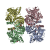
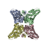
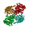
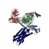
| Title | Crystal structure of the TtnD decarboxylase from the tautomycetin biosynthesis pathway of Streptomyces griseochromogenes with apo form at 1.83 A resolution (I222) | ||||||
 Components Components | UbiD-like decarboxylase | ||||||
 Keywords Keywords | LYASE / TtnD / decarboxylase / tautomycetin biosynthesis / prFMN binding / Structural Genomics / PSI-Biology / Enzyme Discovery for Natural Product Biosynthesis / NatPro | ||||||
| Function / homology |  Function and homology information Function and homology informationpyrrole-2-carboxylate decarboxylase / pyrrole-2-carboxylate decarboxylase activity / ferulate metabolic process / cinnamic acid catabolic process / nucleotide binding / metal ion binding / cytoplasm Similarity search - Function | ||||||
| Biological species |  Streptomyces griseochromogenes (bacteria) Streptomyces griseochromogenes (bacteria) | ||||||
| Method |  X-RAY DIFFRACTION / SYNCHROTRON / MOLECULAR REPLACEMENT / molecular replacement / Resolution: 1.83 Å X-RAY DIFFRACTION / SYNCHROTRON / MOLECULAR REPLACEMENT / molecular replacement / Resolution: 1.83 Å | ||||||
 Authors Authors | Han, L. / Rudolf, J.D. / Chang, C.-Y. / Miller, M.D. / Soman, J. / Phillips Jr., G.N. / Shen, B. / Enzyme Discovery for Natural Product Biosynthesis (NatPro) | ||||||
| Funding support |  United States, 1items United States, 1items
| ||||||
 Citation Citation | Journal: ACS Chem. Biol. / Year: 2018 Title: Biochemical and Structural Characterization of TtnD, a Prenylated FMN-Dependent Decarboxylase from the Tautomycetin Biosynthetic Pathway. Authors: Annaval, T. / Han, L. / Rudolf, J.D. / Xie, G. / Yang, D. / Chang, C.Y. / Ma, M. / Crnovcic, I. / Miller, M.D. / Soman, J. / Xu, W. / Phillips Jr., G.N. / Shen, B. | ||||||
| History |
|
- Structure visualization
| Structure viewer | Molecule: MolmilJmol/JSmol |
|---|
- Downloads & links
-Download
| PDBx/mmCIF format | 6da7.cif.gz | 209.8 KB | Display | PDBx/mmCIF format |
|---|---|---|---|---|
| PDB format | pdb6da7.ent.gz | 163.7 KB | Display | PDB format |
| PDBx/mmJSON format | 6da7.json.gz | Tree view | PDBx/mmJSON format | |
| Others |  Other downloads Other downloads |
-Validation report
| Arichive directory | https://data.pdbj.org/pub/pdb/validation_reports/da/6da7ftp://data.pdbj.org/pub/pdb/validation_reports/da/6da7 | HTTPS FTP |
|---|
-Related structure data
| Related structure data |  6da6SC  6da9C S: Starting model for refinement C: citing same article ( |
|---|---|
| Similar structure data | |
| Other databases |


-Links
 PDBj
PDBj

- Assembly
| Deposited unit | 
| ||||||||||||||||||
|---|---|---|---|---|---|---|---|---|---|---|---|---|---|---|---|---|---|---|---|
| 1 | 
| ||||||||||||||||||
| Unit cell |
| ||||||||||||||||||
| Components on special symmetry positions |
|

-Components
-Protein , 1 types, 1 molecules A
| #1: Protein | Mass: 54783.375 Da / Num. of mol.: 1 Source method: isolated from a genetically manipulated source Source: (gene. exp.) Streptomyces griseochromogenes (bacteria)Gene: ttnD Plasmid details: ttnD gene cloned in pCDFDuet-1 between EcoRI and HindIII sites Plasmid: pBS13033 / Production host: References: UniProt: C6ZCR8, Lyases; Carbon-carbon lyases; Carboxy-lyases |
|---|
-Non-polymers , 5 types, 254 molecules 


| #2: Chemical | ChemComp-NA /  Mass: 22.990 Da / Num. of mol.: 1 / Source method: obtained synthetically / Formula: Na Mass: 22.990 Da / Num. of mol.: 1 / Source method: obtained synthetically / Formula: Na | ||||
|---|---|---|---|---|---|
| #3: Chemical | ChemComp-EPE /  Mass: 238.305 Da / Num. of mol.: 1 / Source method: obtained synthetically / Formula: C8H18N2O4S / Comment: pH buffer*YM Mass: 238.305 Da / Num. of mol.: 1 / Source method: obtained synthetically / Formula: C8H18N2O4S / Comment: pH buffer*YM | ||||
| #4: Chemical | ChemComp-GOL /  Mass: 92.094 Da / Num. of mol.: 18 / Source method: obtained synthetically / Formula: C3H8O3 Mass: 92.094 Da / Num. of mol.: 18 / Source method: obtained synthetically / Formula: C3H8O3#5: Chemical | ChemComp-UNL / | Num. of mol.: 1 / Source method: obtained synthetically #6: Water | ChemComp-HOH / | Mass: 18.015 Da / Num. of mol.: 233 / Source method: isolated from a natural source / Formula: H2O |
-Experimental details
-Experiment
| Experiment | Method: X-RAY DIFFRACTION / Number of used crystals: 1 |
|---|
- Sample preparation
| Crystal | Colour: clear / Density Matthews: 2.66 Å3/Da / Density % sol: 53.68 % | ||||||||||||||||||||||||||||||||||||||||
|---|---|---|---|---|---|---|---|---|---|---|---|---|---|---|---|---|---|---|---|---|---|---|---|---|---|---|---|---|---|---|---|---|---|---|---|---|---|---|---|---|---|
| Crystal grow | Temperature: 298 K / Method: vapor diffusion, sitting drop / pH: 7 Details: 40.0 v/v pentaerythritol propoxylate (5/4 PO/OH); 0.10 M HEPES, pH=7.0; 0.20 M sodium thiocyanate, VAPOR DIFFUSION, SITTING DROP | ||||||||||||||||||||||||||||||||||||||||
| Experiment crystal grow comp |
| ||||||||||||||||||||||||||||||||||||||||
| Experiment crystal grow sol |
| ||||||||||||||||||||||||||||||||||||||||
| Experiment crystal cryo treatment | Cooling details: Direct immersion in liquid nitrogen. Final solution details: 20% glycerol in precipitant solution |
-Data collection
| Diffraction | Mean temperature: 100 K | ||||||||||||||||||||||||||||||||||||||||||||||||||||||||||||||||||||||||||||||||||||||||||||||||||||||||||||||||||||||||||||||||||||||||||||||||||||||||||||||||||||||||||||||||||||||||||||||||||||||||||||||||||||||||||||||||||||||||||||||||||||||||||||
|---|---|---|---|---|---|---|---|---|---|---|---|---|---|---|---|---|---|---|---|---|---|---|---|---|---|---|---|---|---|---|---|---|---|---|---|---|---|---|---|---|---|---|---|---|---|---|---|---|---|---|---|---|---|---|---|---|---|---|---|---|---|---|---|---|---|---|---|---|---|---|---|---|---|---|---|---|---|---|---|---|---|---|---|---|---|---|---|---|---|---|---|---|---|---|---|---|---|---|---|---|---|---|---|---|---|---|---|---|---|---|---|---|---|---|---|---|---|---|---|---|---|---|---|---|---|---|---|---|---|---|---|---|---|---|---|---|---|---|---|---|---|---|---|---|---|---|---|---|---|---|---|---|---|---|---|---|---|---|---|---|---|---|---|---|---|---|---|---|---|---|---|---|---|---|---|---|---|---|---|---|---|---|---|---|---|---|---|---|---|---|---|---|---|---|---|---|---|---|---|---|---|---|---|---|---|---|---|---|---|---|---|---|---|---|---|---|---|---|---|---|---|---|---|---|---|---|---|---|---|---|---|---|---|---|---|---|---|---|---|---|---|---|---|---|---|---|---|---|---|---|---|---|---|
| Diffraction source | Source: SYNCHROTRON / Site: APS / Beamline: 21-ID-F / Wavelength: 0.9787 Å | ||||||||||||||||||||||||||||||||||||||||||||||||||||||||||||||||||||||||||||||||||||||||||||||||||||||||||||||||||||||||||||||||||||||||||||||||||||||||||||||||||||||||||||||||||||||||||||||||||||||||||||||||||||||||||||||||||||||||||||||||||||||||||||
| Detector | Type: RAYONIX MX-225 / Detector: CCD / Date: Mar 28, 2015 | ||||||||||||||||||||||||||||||||||||||||||||||||||||||||||||||||||||||||||||||||||||||||||||||||||||||||||||||||||||||||||||||||||||||||||||||||||||||||||||||||||||||||||||||||||||||||||||||||||||||||||||||||||||||||||||||||||||||||||||||||||||||||||||
| Radiation | Monochromator: Si(111) / Protocol: SINGLE WAVELENGTH / Monochromatic (M) / Laue (L): M / Scattering type: x-ray | ||||||||||||||||||||||||||||||||||||||||||||||||||||||||||||||||||||||||||||||||||||||||||||||||||||||||||||||||||||||||||||||||||||||||||||||||||||||||||||||||||||||||||||||||||||||||||||||||||||||||||||||||||||||||||||||||||||||||||||||||||||||||||||
| Radiation wavelength | Wavelength: 0.9787 Å / Relative weight: 1 | ||||||||||||||||||||||||||||||||||||||||||||||||||||||||||||||||||||||||||||||||||||||||||||||||||||||||||||||||||||||||||||||||||||||||||||||||||||||||||||||||||||||||||||||||||||||||||||||||||||||||||||||||||||||||||||||||||||||||||||||||||||||||||||
| Reflection | Resolution: 1.83→26.77 Å / Num. obs: 52018 / % possible obs: 99.8 % / Redundancy: 3.858 % / Biso Wilson estimate: 27.23 Å2 / CC1/2: 0.999 / Rmerge(I) obs: 0.079 / Rpim(I) all: 0.031 / Rrim(I) all: 0.085 / Χ2: 0.944 / Net I/σ(I): 18.5 | ||||||||||||||||||||||||||||||||||||||||||||||||||||||||||||||||||||||||||||||||||||||||||||||||||||||||||||||||||||||||||||||||||||||||||||||||||||||||||||||||||||||||||||||||||||||||||||||||||||||||||||||||||||||||||||||||||||||||||||||||||||||||||||
| Reflection shell | Diffraction-ID: 1
|
-Phasing
| Phasing | Method: molecular replacement | |||||||||
|---|---|---|---|---|---|---|---|---|---|---|
| Phasing MR |
|
- Processing
| Software |
| |||||||||||||||||||||||||||||||||||||||||||||||||||||||||||||||||||||||||||||||||||||||||||||||||||||||||||||||||||||||||||||
|---|---|---|---|---|---|---|---|---|---|---|---|---|---|---|---|---|---|---|---|---|---|---|---|---|---|---|---|---|---|---|---|---|---|---|---|---|---|---|---|---|---|---|---|---|---|---|---|---|---|---|---|---|---|---|---|---|---|---|---|---|---|---|---|---|---|---|---|---|---|---|---|---|---|---|---|---|---|---|---|---|---|---|---|---|---|---|---|---|---|---|---|---|---|---|---|---|---|---|---|---|---|---|---|---|---|---|---|---|---|---|---|---|---|---|---|---|---|---|---|---|---|---|---|---|---|---|
| Refinement | Method to determine structure: MOLECULAR REPLACEMENT Starting model: 6DA6 Resolution: 1.83→26.77 Å / Cor.coef. Fo:Fc: 0.954 / Cor.coef. Fo:Fc free: 0.944 / SU R Cruickshank DPI: 0.152 / Cross valid method: THROUGHOUT / σ(F): 0 / SU R Blow DPI: 0.118 / SU Rfree Blow DPI: 0.109 / SU Rfree Cruickshank DPI: 0.108 Details: 1. TLS GROUPS WERE ASSIGNED WITH THE AID OF TLSMD. 2. ZERO OCCUPANCY HYDROGENS IN THEIR RIDING POSITION WERE USED AS ANTI-BUMPING RESTRAINTS. 3. HEPES FROM THE CRYSTALLIZATION BUFFER IS ...Details: 1. TLS GROUPS WERE ASSIGNED WITH THE AID OF TLSMD. 2. ZERO OCCUPANCY HYDROGENS IN THEIR RIDING POSITION WERE USED AS ANTI-BUMPING RESTRAINTS. 3. HEPES FROM THE CRYSTALLIZATION BUFFER IS BOUND IN THE pr-FMN BINDING SITE. 4. AN UNKNOWN LIGAND (UNL) IS MODELED. THERE ARE 2 UNL'S BOUND IN THE TETRAMER. THE SHAPE LOOKS LIKE BENZOATE OR NIACIN. THE SITE IS ON A CRYSTALLOGRPAHIC TWO-FOLD AXIS.
| |||||||||||||||||||||||||||||||||||||||||||||||||||||||||||||||||||||||||||||||||||||||||||||||||||||||||||||||||||||||||||||
| Displacement parameters | Biso max: 166.49 Å2 / Biso mean: 44.26 Å2 / Biso min: 14.42 Å2
| |||||||||||||||||||||||||||||||||||||||||||||||||||||||||||||||||||||||||||||||||||||||||||||||||||||||||||||||||||||||||||||
| Refine analyze | Luzzati coordinate error obs: 0.25 Å | |||||||||||||||||||||||||||||||||||||||||||||||||||||||||||||||||||||||||||||||||||||||||||||||||||||||||||||||||||||||||||||
| Refinement step | Cycle: final / Resolution: 1.83→26.77 Å
| |||||||||||||||||||||||||||||||||||||||||||||||||||||||||||||||||||||||||||||||||||||||||||||||||||||||||||||||||||||||||||||
| Refine LS restraints |
| |||||||||||||||||||||||||||||||||||||||||||||||||||||||||||||||||||||||||||||||||||||||||||||||||||||||||||||||||||||||||||||
| LS refinement shell | Resolution: 1.83→1.88 Å / Rfactor Rfree error: 0 / Total num. of bins used: 20
| |||||||||||||||||||||||||||||||||||||||||||||||||||||||||||||||||||||||||||||||||||||||||||||||||||||||||||||||||||||||||||||
| Refinement TLS params. | Method: refined / Refine-ID: X-RAY DIFFRACTION
| |||||||||||||||||||||||||||||||||||||||||||||||||||||||||||||||||||||||||||||||||||||||||||||||||||||||||||||||||||||||||||||
| Refinement TLS group |
|
