 Movie
Movie Controller

[English] 日本語
 Yorodumi
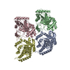
- PDB-6zn2: Partial structure of tyrosine hydroxylase in complex with dopamin... -
+ Open data

- Basic information
| Entry | Database: PDB / ID: 6zn2 | |||||||||
|---|---|---|---|---|---|---|---|---|---|---|
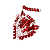
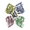
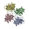
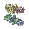
| Title | Partial structure of tyrosine hydroxylase in complex with dopamine showing the catalytic domain and an alpha-helix from the regulatory domain involved in dopamine binding. | |||||||||
 Components Components |
| |||||||||
 Keywords Keywords | OXIDOREDUCTASE / Tetramer / Dopamine / Catecholamine / Brain / Parkinson | |||||||||
| Function / homology |  Function and homology information Function and homology informationtyrosine 3-monooxygenase / tyrosine 3-monooxygenase activity / dopamine biosynthetic process from tyrosine / embryonic camera-type eye morphogenesis / norepinephrine biosynthetic process / epinephrine biosynthetic process / serotonin biosynthetic process / Catecholamine biosynthesis / hyaloid vascular plexus regression / eye photoreceptor cell development ...tyrosine 3-monooxygenase / tyrosine 3-monooxygenase activity / dopamine biosynthetic process from tyrosine / embryonic camera-type eye morphogenesis / norepinephrine biosynthetic process / epinephrine biosynthetic process / serotonin biosynthetic process / Catecholamine biosynthesis / hyaloid vascular plexus regression / eye photoreceptor cell development / melanosome membrane / mating behavior / dopamine biosynthetic process / eating behavior / regulation of heart contraction / pigmentation / smooth endoplasmic reticulum / synaptic transmission, dopaminergic / anatomical structure morphogenesis / heart morphogenesis / visual perception / animal organ morphogenesis / learning / locomotory behavior / cognition / memory / cytoplasmic side of plasma membrane / heart development / synaptic vesicle / cytoplasmic vesicle / response to ethanol / perikaryon / response to hypoxia / neuron projection / iron ion binding / axon / perinuclear region of cytoplasm / enzyme binding / identical protein binding / nucleus / cytosol / cytoplasm Similarity search - Function | |||||||||
| Biological species |  Homo sapiens (human) Homo sapiens (human) | |||||||||
| Method | ELECTRON MICROSCOPY / single particle reconstruction / cryo EM / Resolution: 4.3 Å | |||||||||
 Authors Authors | Bueno-Carrasco, M.T. / Cuellar, J. / Santiago, C. / Valpuesta, J.M. / Martinez, A. / Flydal, M.I. | |||||||||
| Funding support |  Spain, 2items Spain, 2items
| |||||||||
 Citation Citation | Journal: Nat Commun / Year: 2022 Title: Structural mechanism for tyrosine hydroxylase inhibition by dopamine and reactivation by Ser40 phosphorylation. Authors: María Teresa Bueno-Carrasco / Jorge Cuéllar / Marte I Flydal / César Santiago / Trond-André Kråkenes / Rune Kleppe / José R López-Blanco / Miguel Marcilla / Knut Teigen / Sara Alvira ...Authors: María Teresa Bueno-Carrasco / Jorge Cuéllar / Marte I Flydal / César Santiago / Trond-André Kråkenes / Rune Kleppe / José R López-Blanco / Miguel Marcilla / Knut Teigen / Sara Alvira / Pablo Chacón / Aurora Martinez / José M Valpuesta /   Abstract: Tyrosine hydroxylase (TH) catalyzes the rate-limiting step in the biosynthesis of dopamine (DA) and other catecholamines, and its dysfunction leads to DA deficiency and parkinsonisms. Inhibition by ...Tyrosine hydroxylase (TH) catalyzes the rate-limiting step in the biosynthesis of dopamine (DA) and other catecholamines, and its dysfunction leads to DA deficiency and parkinsonisms. Inhibition by catecholamines and reactivation by S40 phosphorylation are key regulatory mechanisms of TH activity and conformational stability. We used Cryo-EM to determine the structures of full-length human TH without and with DA, and the structure of S40 phosphorylated TH, complemented with biophysical and biochemical characterizations and molecular dynamics simulations. TH presents a tetrameric structure with dimerized regulatory domains that are separated 15 Å from the catalytic domains. Upon DA binding, a 20-residue α-helix in the flexible N-terminal tail of the regulatory domain is fixed in the active site, blocking it, while S40-phosphorylation forces its egress. The structures reveal the molecular basis of the inhibitory and stabilizing effects of DA and its counteraction by S40-phosphorylation, key regulatory mechanisms for homeostasis of DA and TH. | |||||||||
| History |
|
- Structure visualization
| Movie |
Movie viewer |
|---|---|
| Structure viewer | Molecule: MolmilJmol/JSmol |
- Downloads & links
-Download
| PDBx/mmCIF format | 6zn2.cif.gz | 333.3 KB | Display | PDBx/mmCIF format |
|---|---|---|---|---|
| PDB format | pdb6zn2.ent.gz | 232.3 KB | Display | PDB format |
| PDBx/mmJSON format | 6zn2.json.gz | Tree view | PDBx/mmJSON format | |
| Others |  Other downloads Other downloads |
-Validation report
| Arichive directory | https://data.pdbj.org/pub/pdb/validation_reports/zn/6zn2ftp://data.pdbj.org/pub/pdb/validation_reports/zn/6zn2 | HTTPS FTP |
|---|
-Related structure data
| Related structure data |  11309MC  6zvpC  6zzuC  7a2gC  7pimC M: map data used to model this data C: citing same article ( |
|---|---|
| Similar structure data |


-Links
 PDBj
PDBj
- Assembly
| Deposited unit | 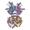
| |||||||||||||||||||||||||||||||||||||||||||||||||||||||||||||||||||||||||||||||||||||||||||||||||||||||||||||||||||||||||||||||||||||||||||||||||||||
|---|---|---|---|---|---|---|---|---|---|---|---|---|---|---|---|---|---|---|---|---|---|---|---|---|---|---|---|---|---|---|---|---|---|---|---|---|---|---|---|---|---|---|---|---|---|---|---|---|---|---|---|---|---|---|---|---|---|---|---|---|---|---|---|---|---|---|---|---|---|---|---|---|---|---|---|---|---|---|---|---|---|---|---|---|---|---|---|---|---|---|---|---|---|---|---|---|---|---|---|---|---|---|---|---|---|---|---|---|---|---|---|---|---|---|---|---|---|---|---|---|---|---|---|---|---|---|---|---|---|---|---|---|---|---|---|---|---|---|---|---|---|---|---|---|---|---|---|---|---|---|
| 1 |
| |||||||||||||||||||||||||||||||||||||||||||||||||||||||||||||||||||||||||||||||||||||||||||||||||||||||||||||||||||||||||||||||||||||||||||||||||||||
| Noncrystallographic symmetry (NCS) | NCS domain:
NCS domain segments:
NCS ensembles :
NCS oper:
|
-Components
| #1: Protein | Mass: 38087.766 Da / Num. of mol.: 4 Source method: isolated from a genetically manipulated source Source: (gene. exp.) Homo sapiens (human) / Gene: TH, TYH / Production host:  #2: Protein/peptide | Mass: 1874.081 Da / Num. of mol.: 4 Source method: isolated from a genetically manipulated source Source: (gene. exp.) Homo sapiens (human) / Production host: #3: Chemical | ChemComp-LDP /   Mass: 153.178 Da / Num. of mol.: 4 / Source method: obtained synthetically / Formula: C8H11NO2 / Comment: medication*YM Mass: 153.178 Da / Num. of mol.: 4 / Source method: obtained synthetically / Formula: C8H11NO2 / Comment: medication*YM#4: Chemical | ChemComp-FE /   Mass: 55.845 Da / Num. of mol.: 4 / Source method: obtained synthetically / Formula: Fe Mass: 55.845 Da / Num. of mol.: 4 / Source method: obtained synthetically / Formula: FeHas ligand of interest | N | |
|---|
-Experimental details
-Experiment
| Experiment | Method: ELECTRON MICROSCOPY |
|---|---|
| EM experiment | Aggregation state: PARTICLE / 3D reconstruction method: single particle reconstruction |
- Sample preparation
| Component | Name: Tyrosine Hydroxylase / Type: COMPLEX / Entity ID: #1-#2 / Source: RECOMBINANT |
|---|---|
| Source (natural) | Organism: Homo sapiens (human) |
| Source (recombinant) | Organism: |
| Buffer solution | pH: 7.4 |
| Specimen | Embedding applied: NO / Shadowing applied: NO / Staining applied: NO / Vitrification applied: YES |
| Vitrification | Cryogen name: ETHANE |
- Electron microscopy imaging
| Experimental equipment |  Model: Titan Krios / Image courtesy: FEI Company |
|---|---|
| Microscopy | Model: FEI TITAN KRIOS |
| Electron gun | Electron source:  FIELD EMISSION GUN / Accelerating voltage: 300 kV / Illumination mode: OTHER FIELD EMISSION GUN / Accelerating voltage: 300 kV / Illumination mode: OTHER |
| Electron lens | Mode: BRIGHT FIELD |
| Image recording | Electron dose: 1 e/Å2 / Film or detector model: GATAN K2 SUMMIT (4k x 4k) |
- Processing
| Software |
| ||||||||||||||||||||||||||||||||||||||||||
|---|---|---|---|---|---|---|---|---|---|---|---|---|---|---|---|---|---|---|---|---|---|---|---|---|---|---|---|---|---|---|---|---|---|---|---|---|---|---|---|---|---|---|---|
| CTF correction | Type: NONE | ||||||||||||||||||||||||||||||||||||||||||
| 3D reconstruction | Resolution: 4.3 Å / Resolution method: FSC 0.143 CUT-OFF / Num. of particles: 125033 / Symmetry type: POINT | ||||||||||||||||||||||||||||||||||||||||||
| Refinement | Cross valid method: NONE Stereochemistry target values: GeoStd + Monomer Library + CDL v1.2 | ||||||||||||||||||||||||||||||||||||||||||
| Displacement parameters | Biso mean: 259.35 Å2 | ||||||||||||||||||||||||||||||||||||||||||
| Refine LS restraints |
| ||||||||||||||||||||||||||||||||||||||||||
| Refine LS restraints NCS |
|
